. 2024 Apr 27;14(1):9700.
doi: 10.1038/s41598-024-60540-6.

Sphingosine-1-phosphate receptor 1/5 selective agonist alleviates ocular vascular pathologies

Affiliations

Sphingosine-1-phosphate receptor 1/5 selective agonist alleviates ocular vascular pathologies

Shinsuke Nakamura et al. Sci Rep. .

Abstract

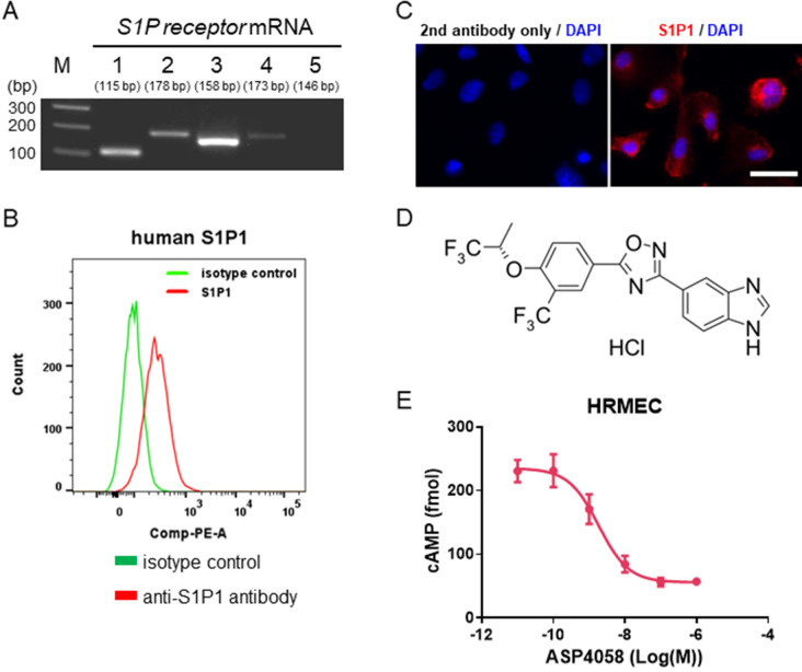
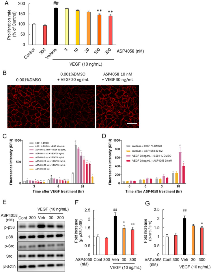
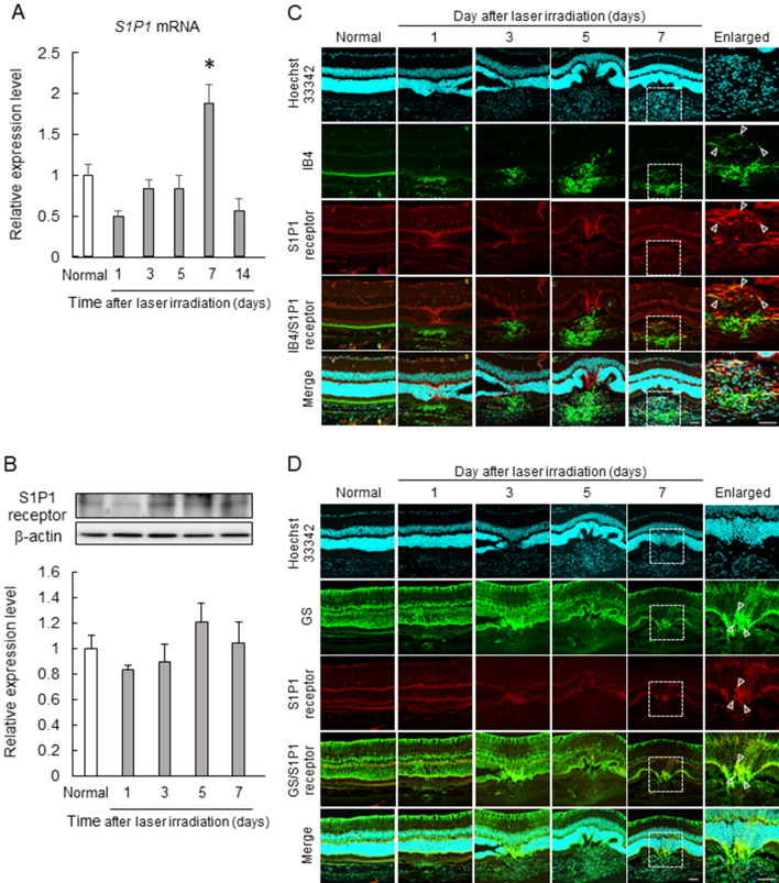
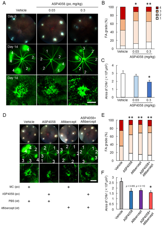
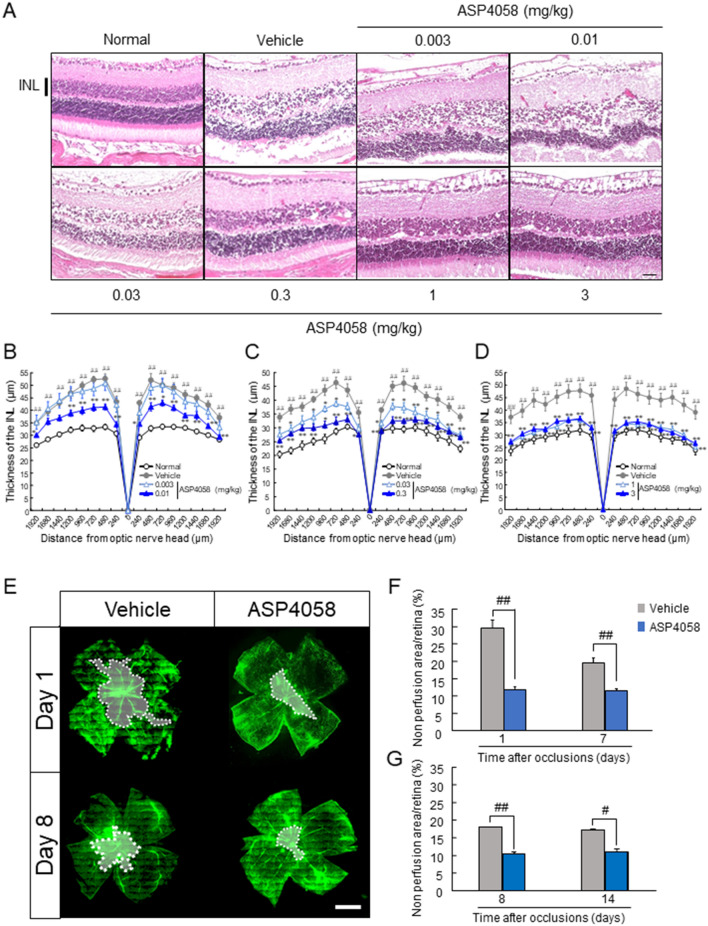
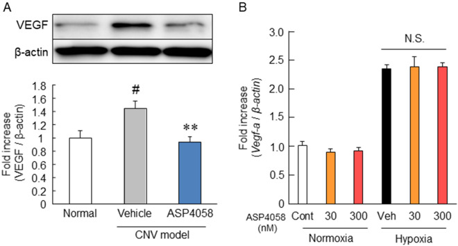
Ocular abnormal angiogenesis and edema are featured in several ocular diseases. S1P signaling via S1P1 likely is part of the negative feedback mechanism necessary to maintain vascular health. In this study, we conducted pharmacological experiments to determine whether ASP4058, a sphingosine 1-phosphate receptor 1/5 (S1P1/5) agonist, is useful in abnormal vascular pathology in the eye. First, human retinal microvascular endothelial cells (HRMECs) were examined using vascular endothelial growth factor (VEGF)-induced cell proliferation and hyperpermeability. ASP4058 showed high affinity and inhibited VEGF-induced proliferation and hyperpermeability of HRMECs. Furthermore, S1P1 expression and localization changes were examined in the murine laser-induced choroidal neovascularization (CNV) model, a mouse model of exudative age-related macular degeneration, and the efficacy of ASP4058 was verified. In the CNV model mice, S1P1 tended to decrease in expression immediately after laser irradiation and colocalized with endothelial cells and Müller glial cells. Oral administration of ASP4058 also suppressed vascular hyperpermeability and CNV, and the effect was comparable to that of the intravitreal administration of aflibercept, an anti-VEGF drug. Next, efficacy was also examined in a retinal vein occlusion (RVO) model in which retinal vascular permeability was increased. ASP4058 dose-dependently suppressed the intraretinal edema. In addition, it suppressed the expansion of the perfusion area observed in the RVO model. ASP4058 also suppressed the production of VEGF in the eye. Collectively, ASP4058 can be a potential therapeutic agent that normalizes abnormal vascular pathology, such as age-related macular degeneration and RVO, through its direct action on endothelial cells.


Conflict of interest statement

S.N., M.S., S.N., and H.H. received a research grant from Astellas Pharma Inc. R.Y., T.M., and S.K. are employees of Astellas Pharma Inc. Y.I. and M.G. are employees of Astellas Institute for Regenerative Medicine. H.Y., A.N., and K.T. declare no competing interests.

Figures

Figure 1
Figure 1
ASP4058 functions as an S1P1 agonist in HRMEC. ASP4058, an S1P1 and S1P5 selective agonist, exerts agonistic effects on HRMEC via S1P1. (A) RT-PCR analysis showing all S1P receptor subtypes mRNA expression except for S1P5 in HRMECs. (B) Flow cytometry analysis showing the S1P1 cell surface expression in HRMECs. Data are shown as a histogram overlay: isotype control-stained cells (green) and anti-S1P1 antibody-stained cells (red). (C) The representative immunofluorescence image of S1P1 receptor (red) and DAPI (blue) of HRMECs. Scale bar = 30 µm. (D) Chemical structure of ASP4058. (E) ASP4058 inhibited forskolin-induced cAMP accumulation in HRMECs. Data are presented as the mean ± SEM (n = 5).
Figure 2
Figure 2
ASP4058 attenuated HRMEC proliferation and hyperpermeability induced by VEGF. ASP4058 reduced HRMECs proliferation and hyperpermeability induced by VEGF. (A) ASP4058 decreased VEGF-induced HRMECs proliferation concentration-dependently. Data are shown as the mean ± SEM (n = 6). **; p < 0.01 vs. vehicle-treated group, ##; p < 0.01 vs. control group (Tukey’s test). (B) Representative immunofluorescence micrographs analyzing VE-cadherin expression and localization in the VEGF-stimulated HRMEC monolayer. HRMEC monolayers were left untreated (left panel) or treated with 30 ng/mL VEGF (middle and right panel) after pretreatment with 0.001% DMSO (left and middle panel) or 10 nM ASP4058 (right panel) and stained with antibody against VE-cadherin. Scale bar = 50 µm. (C, D) Pre- or post-treatment with ASP4058 inhibited VEGF-induced hyperpermeability. HRMEC monolayer cultured on Transwell inserts were permeabilized by treatment with 30 ng/mL VEGF, and measured for permeability by diffusing FITC-conjugated dextran (500 KDa) across the insert. (C) Treatment with ASP4058 1 h before VEGF treatment inhibited the increase of permeability induced by VEGF. RFU, relative fluorescent units. Data are shown as the mean ± SEM (n = 5). *; p < 0.05, **; p < 0.01 vs. 0.001% DMSO + 30 ng/mL VEGF group (Dunnett’s test), #; p < 0.05, ##; p < 0.01 vs. 0.001% DMSO group (Student’s t-test). (D) ASP4058 post-treatment from 3 h after VEGF treatment suppressed VEGF-induced hyperpermeability. Data are presented as the mean ± SEM (n = 6). #; p < 0.05, ##; p < 0.01 vs. 30 ng/mL VEGF + 0.001% DMSO group (Student’s t-test). (EG) ASP4058 treatment 1 h before VEGF treatment inhibited VEGF-induced phosphorylation of p38 (F) and Src (G). Data are shown as the mean ± SEM (n = 6). ##; p < 0.01 vs. Vehicle-treated group, *; p < 0.05, **; p < 0.01 vs. Control group (Tukey’s test).
Figure 3
Figure 3
Expression and localization of S1P1 in the laser-induced CNV model. CNV induction affected the expression level and localization of S1P1. The expression level of S1p1 mRNA at one, three, five, seven, and 14 days after laser irradiation (A) and S1P1 protein at one, three, five, and seven days after laser irradiation (B) in the RPE-choroid complex. Data are presented as the mean ± SEM (n = 5–10). *; p < 0.05 vs. normal group (Games–Howell post-hoc test). (C, D) Immunohistochemistry of Hoechst 33,342 (cyan), S1P1 (red), IB4 (green) (C), and GS (green) (D); insets are enlarged image of the enclosed area. Scale bars show 50 µm.
Figure 4
Figure 4
Anti-angiogenic effects of ASP4058 on the CNV model mice. ASP4058 showed anti-angiogenic effects on CNV development in the laser-induced CNV model. (AC) The laser-induced CNV model mice were orally administered ASP4058 (0.03 or 0.3 mg/kg) once a day. (A) Representative fundus images immediately after laser irradiation (day zero) and after fluorescein injection (day 14) with grades (1–4) and CNV lesion visualized by FITC-dextran. (B) Quantitative data of vascular leakages (grade 1–4) of each spot. **; p < 0.01 vs. vehicle-administered group (Kruskal–Wallis test). (C) Quantification of the mean size of CNVs. The scale bar indicates 100 µm. Data are presented as the mean ± SEM (n = 11 or 12). **; p < 0.01 vs. vehicle-administered group (Dunnett’s test). (DF) Murine laser-induced CNV model were orally administered 0.3 mg/kg ASP4058 once a day and/or intravitreal administered anti-VEGF agent, aflibercept. (D) Representative fundus images immediately after laser irradiation (day zero) and after fluorescein injection (day 14) with grades and the CNV lesion. (E) Quantitative data of vascular leakages (grade 1–4) of each spot. **; p < 0.01 vs. vehicle-administered group (Kruskal–Wallis test). (F) Quantification of the mean size of CNVs. The scale bar shows 100 µm. Data are presented as the mean ± SEM (n = 11–14). *; p < 0.05 vs. vehicle-administered group (Tukey’s test).
Figure 5
Figure 5
Effect of ASP4058 on the formation of retina edema and non-perfused region in the RVO model mice. ASP4058 suppressed the formation of retinal edema and non-perfusion area in the RVO model. (A) Photomicrographs of representative H&E-stained retinal sections of normal, vehicle, and ASP4058 groups at 0.003, 0.01, 0.03, 0.3, 1, and 3 mg/kg orally administered just after laser irradiation. Images were taken at 500 µm from the optic nerve head. Scale bar indicates 50 µm. (BD) Quantitative data of the thickness of the INL. Each graph contains 0.003 and 0.01 (B), 0.03 and 0.3 (C), and 1 and 3 mg/kg (D) ASP4058. Data are presented as the mean ± SEM (n = 10). *; p < 0.05, **; p < 0.01 vs. vehicle-treated group (Dunnett’s T3 test), ##; p < 0.01 vs. normal group (Welch’s t-test). (EG) The effect of ASP4058 at 0.3 mg/kg on the formation of a non-perfusion area. ASP4058 was orally administered 12 h before and just after laser irradiation (early phase) or twice seven days after occlusion (late phase). To examine the effect of ASP4058 treatment in the early and late phase, the retinas were collected at days one or seven, and eight or 14, respectively. (E) Representative images of the non-perfusion area on days one and eight. Scale bar shows 500 µm. (FG) Quantitative data of the ratio of the non-perfusion area. ASP4058 treatment in the early phase (F) and late phase (G) inhibited the formation of the non-perfusion region. Data are presented as the mean ± SEM (n = 7–10). #; p < 0.05, ##; p < 0.01 vs. vehicle-administered group (Welch’s t-test).
Figure 6
Figure 6
Effects of ASP4058 on VEGF production in in vivo and in vitro models. ASP4058 reduced the VEGF expression level in the CNV model mice. (A) Western immunoblotting analysis of VEGF and β-actin in the RPE-choroid complex five days after the laser irradiation. ASP4058 was orally administered once a day for six days from one day before laser irradiation. Data are presented as the mean ± SEM (n = 6–8). **; p < 0.01 vs. vehicle-administered group, #; p < 0.05 vs. normal group (Tukey’s test). (B) The presence or absence of the inhibitory effect of ASP4058 on VEGF mRNA expression was examined using RT-PCR. Pre-treatment of ASP4058 (final concentration: 30 or 300 nM) was started 1 h before hypoxia induction of treatment. Data are shown as the mean ± SEM (n = 6). N.S.; p > 0.05 vs. vehicle-treated group (Tukey’s test).

Similar articles

Cited by

References

    1. Campochiaro PA. Ocular neovascularization. J. Mol. Med. (Berl.) 2013;91:311–321. doi: 10.1007/s00109-013-0993-5. - DOI - PMC - PubMed
    1. Rajappa M, Saxena P, Kaur J. Ocular angiogenesis: Mechanisms and recent advances in therapy. Adv. Clin. Chem. 2010;50:103–121. doi: 10.1016/S0065-2423(10)50006-4. - DOI - PubMed
    1. Nguyen DH, Luo J, Zhang K, Zhang M. Current therapeutic approaches in neovascular age-related macular degeneration. Discov. Med. 2013;15:343–348. - PubMed
    1. Falavarjani KG, Nguyen QD. Adverse events and complications associated with intravitreal injection of anti-VEGF agents: A review of literature. Eye (Lond.) 2013;27:787–794. doi: 10.1038/eye.2013.107. - DOI - PMC - PubMed
    1. Maguire MG, et al. Five-year outcomes with anti-vascular endothelial growth factor treatment of neovascular age-related macular degeneration: The comparison of age-related macular degeneration treatments trials. Ophthalmology. 2016;123:1751–1761. doi: 10.1016/j.ophtha.2016.03.045. - DOI - PMC - PubMed

Publication types

MeSH terms

Substances