

Electrospun nanofibers synthesized from polymers incorporated with bioactive compounds for wound healing
- PMID: 38678271
- PMCID: PMC11056076
- DOI: 10.1186/s12951-024-02491-8
Electrospun nanofibers synthesized from polymers incorporated with bioactive compounds for wound healing
Abstract
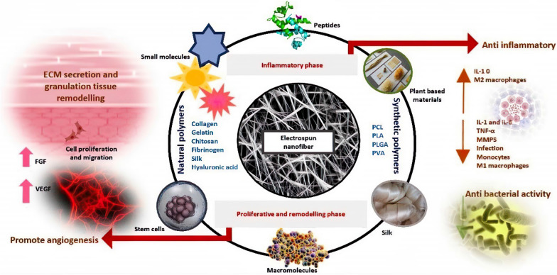
The development of innovative wound dressing materials is crucial for effective wound care. It's an active area of research driven by a better understanding of chronic wound pathogenesis. Addressing wound care properly is a clinical challenge, but there is a growing demand for advancements in this field. The synergy of medicinal plants and nanotechnology offers a promising approach to expedite the healing process for both acute and chronic wounds by facilitating the appropriate progression through various healing phases. Metal nanoparticles play an increasingly pivotal role in promoting efficient wound healing and preventing secondary bacterial infections. Their small size and high surface area facilitate enhanced biological interaction and penetration at the wound site. Specifically designed for topical drug delivery, these nanoparticles enable the sustained release of therapeutic molecules, such as growth factors and antibiotics. This targeted approach ensures optimal cell-to-cell interactions, proliferation, and vascularization, fostering effective and controlled wound healing. Nanoscale scaffolds have significant attention due to their attractive properties, including delivery capacity, high porosity and high surface area. They mimic the Extracellular matrix (ECM) and hence biocompatible. In response to the alarming rise of antibiotic-resistant, biohybrid nanofibrous wound dressings are gradually replacing conventional antibiotic delivery systems. This emerging class of wound dressings comprises biopolymeric nanofibers with inherent antibacterial properties, nature-derived compounds, and biofunctional agents. Nanotechnology, diminutive nanomaterials, nanoscaffolds, nanofibers, and biomaterials are harnessed for targeted drug delivery aimed at wound healing. This review article discusses the effects of nanofibrous scaffolds loaded with nanoparticles on wound healing, including biological (in vivo and in vitro) and mechanical outcomes.
Keywords: Antibacterial; Electrospining; Nanofibrous mats; Nanomaterial; Wound dressing.
© 2024. The Author(s).
Conflict of interest statement
The authors declare that they have no conflict of interest.
Figures


References
-
- Mahmoudi N, Eslahi N, Mehdipour A, Mohammadi M, Akbari M, Samadikuchaksaraei A, et al. Temporary skin grafts based on hybrid graphene oxide-natural biopolymer nanofibers as effective wound healing substitutes: pre-clinical and pathological studies in animal models. J Mater Sci Mater Med. 2017 doi: 10.1007/s10856-017-5874-y. - DOI - PubMed
Publication types
MeSH terms
Substances
LinkOut - more resources
Full Text Sources
Medical
