. 2024 Oct;168(4):e121-e132.
doi: 10.1016/j.jtcvs.2024.03.001. Epub 2024 Apr 27.

Transient receptor potential vanilloid 4 channel inhibition attenuates lung ischemia-reperfusion injury in a porcine lung transplant model

Affiliations

Transient receptor potential vanilloid 4 channel inhibition attenuates lung ischemia-reperfusion injury in a porcine lung transplant model

Raymond J Strobel et al. J Thorac Cardiovasc Surg. 2024 Oct.

Abstract

Objective: Transient receptor potential vanilloid 4 (TRPV4) is a nonselective cation channel important in many physiological and pathophysiological processes, including pulmonary disease. Using a murine model, we previously demonstrated that TRPV4 mediates lung ischemia-reperfusion injury, the major cause of primary graft dysfunction after transplant. The current study tests the hypothesis that treatment with a TRPV4 inhibitor will attenuate lung ischemia-reperfusion injury in a clinically relevant porcine lung transplant model.

Methods: A porcine left-lung transplant model was used. Animals were randomized to 2 treatment groups (n = 5/group): vehicle or GSK2193874 (selective TRPV4 inhibitor). Donor lungs underwent 30 minutes of warm ischemia and 24 hours of cold preservation before left lung allotransplantation and 4 hours of reperfusion. Vehicle or GSK2193874 (1 mg/kg) was administered to the recipient as a systemic infusion after recipient lung explant. Lung function, injury, and inflammatory biomarkers were compared.

Results: After transplant, left lung oxygenation was significantly improved in the TRPV4 inhibitor group after 3 and 4 hours of reperfusion. Lung histology scores and edema were significantly improved, and neutrophil infiltration was significantly reduced in the TRPV4 inhibitor group. TRPV4 inhibitor-treated recipients had significantly reduced expression of interleukin-8, high mobility group box 1, P-selectin, and tight junction proteins (occludin, claudin-5, and zonula occludens-1) in bronchoalveolar lavage fluid as well as reduced angiopoietin-2 in plasma, all indicative of preservation of endothelial barrier function.

Conclusions: Treatment of lung transplant recipients with TRPV4 inhibitor significantly improves lung function and attenuates ischemia-reperfusion injury. Thus, selective TRPV4 inhibition may be a promising therapeutic strategy to prevent primary graft dysfunction after transplant.

Keywords: TRPV4 channel; endothelial barrier; inflammation; ischemia-reperfusion injury; lung transplantation; primary graft dysfunction.


Conflict of interest statement

Conflict of Interest Statement The authors reported no conflicts of interest. The Journal policy requires editors and reviewers to disclose conflicts of interest and to decline handling or reviewing manuscripts for which they may have a conflict of interest. The editors and reviewers of this article have no conflicts of interest.

Figures
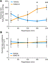
Figure 1.
Figure 1.. Lung function is improved during reperfusion in recipient animals treated with GSK219.
A) Left lung-specific oxygenation (pulmonary vein PaO2/FiO2 ratio) was significantly improved in the GSK219-treated recipient animals vs. vehicle controls after 3 and 4 hours of reperfusion. *P = 0.026, **P = 0.003. B) Dynamic lung compliance was similar between groups. Measurements are at 15 min and then hourly during reperfusion. N = 5/group. Data are expressed as mean ± standard deviation.
Figure 2.
Figure 2.. Lung wet/dry weight ratios after transplant and reperfusion.
No significant differences in wet/dry weight ratios were observed between groups in either left upper lobe, left lingula, or left lower lobe. Data are presented using box-and-whisker plots where the lower and upper borders of the box represent the lower and upper quartiles (25th percentile and 75th percentile); the middle horizontal line represents the median; and the lower and upper whiskers represent the minimum and maximum data values. ns = not significant.
Figure 3.
Figure 3.. Histologic lung injury severity scores were improved in recipient animals treated with GSK219.
A) Histologic scores for alveolar edema and polymorphonuclear neutrophils per high-powered field (PMNs/HPF), as well as composite score, were significantly improved after reperfusion in GSK219-treated animals versus vehicle. Data are presented using box-and-whisker plots where the lower and upper borders of the box represent the lower and upper quartiles (25th percentile and 75th percentile); the middle horizontal line represents the median; and the lower and upper whiskers represent the minimum and maximum data values. *P = 0.048, **P = 0.028, ***P = 0.041, ns = not significant. B) Representative H&E-stained lung sections after 4 hours of reperfusion in vehicle- and GSK219-treated recipient animals (20X magnification).
Figure 4.
Figure 4.. Treatment of recipient animals with GSK219 attenuated neutrophil infiltration in transplanted lungs.
(A) Neutrophil numbers in bronchoalveolar lavage (BAL) fluid measured by differential cell count (% total count) were significantly reduced by GSK219 treatment versus vehicle. (B) The number of neutrophils per high-powered field (HPF), as measured by immunohistochemical staining of lung sections, were significantly reduced by GSK219 treatment versus vehicle. Quantification is shown on the left, and representative images (20X magnification) are shown on the right where neutrophils are stained red. Data are presented using box-and-whisker plots where the lower and upper borders of the box represent the lower and upper quartiles (25th percentile and 75th percentile); the middle horizontal line represents the median; and the lower and upper whiskers represent the minimum and maximum data values. *P = 0.004, **P < 0.001.
Figure 5.
Figure 5.. Treatment of recipient animals with GSK219 attenuated expression of endothelial cell adhesion molecules in lungs after reperfusion.
Expression of P-selectin in BAL fluid was significantly reduced in GSK219-treated animals versus vehicle (A). No significant changes were detected in expression of VCAM1 or ICAM-1 (B,C), which trended lower in GSK219-treated animals. Data are presented using box-and-whisker plots where the lower and upper borders of the box represent the lower and upper quartiles (25th percentile and 75th percentile); the middle horizontal line represents the median; and the lower and upper whiskers represent the minimum and maximum data values. *P = 0.0075.
Figure 6.
Figure 6.. Treatment of recipient animals with GSK219 reduced tight junction protein levels in BAL fluid after reperfusion.
Concentrations of tight junction proteins occludin (A), claudin-5 (B), and ZO-1 (C) in bronchoalveolar lavage (BAL) fluid were significantly reduced in recipient animals treated with GSK219 versus vehicle. The expression of ZO-2 in BAL fluid (D) was not significantly different but trended lower. Data are presented using box-and-whisker plots where the lower and upper borders of the box represent the lower and upper quartiles (25th percentile and 75th percentile); the middle horizontal line represents the median; and the lower and upper whiskers represent the minimum and maximum data values. *P < 0.05.
Figure 7.
Figure 7.. Treatment of recipient animals with GSK219 reduced expression of inflammatory markers after reperfusion.
Expression of HMGB1 in bronchoalveolar lavage (BAL) fluid (A) and Ang-2 in plasma (B) were significantly reduced in animals treated with GSK219 versus vehicle. Expression of IL-8 (C), but not IL-1β (D), was significantly reduced in BAL fluid of GSK219-treated animals. Data are presented using box-and-whisker plots where the lower and upper borders of the box represent the lower and upper quartiles (25th percentile and 75th percentile); the middle horizontal line represents the median; and the lower and upper whiskers represent the minimum and maximum data values. *P < 0.03, **P = 0.001.
Figure 8.
Figure 8.
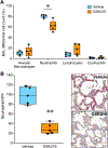
Graphical Abstract detailing methods (top), results (middle), and implications of the study (bottom).

References

    1. Gelman AE, Fisher AJ, Huang HJ, Baz MA, Shaver CM, Egan TM, Mulligan MS. Report of the ISHLT working group on primary lung graft dysfunction Part III: Mechanisms: A 2016 consensus group statement of the International Society for Heart and Lung Transplantation. J Heart Lung Transplant 2017;36(10):1114–1120. doi: 10.1016/j.healun.2017.07.014 - DOI - PMC - PubMed
    1. Laubach VE, Sharma AK. Mechanisms of lung ischemia-reperfusion injury. Curr Opin Organ Transplant 2016;21(3):246–252. doi: 10.1097/MOT.0000000000000304 - DOI - PMC - PubMed
    1. Shah RJ, Diamond JM. Primary Graft Dysfunction (PGD) Following Lung Transplantation. Semin Respir Crit Care Med 2018;39(2):148–154. doi: 10.1055/s-0037-1615797 - DOI - PubMed
    1. Christie JD, Kotloff RM, Ahya VN, Tino G, Pochettino A, Gaughan C, et al. The effect of primary graft dysfunction on survival after lung transplantation. Am J Respir Crit Care Med 2005;171(11):1312–1316. doi: 10.1164/rccm.200409-1243OC - DOI - PMC - PubMed
    1. Daud SA, Yusen RD, Meyers BF, Chakinala MM, Walter MJ, Aloush AA, et al. Impact of immediate primary lung allograft dysfunction on bronchiolitis obliterans syndrome. Am J Respir Crit Care Med 2007;175(5):507–513. doi: 10.1164/rccm.200608-1079OC - DOI - PubMed