. 2024 May 28;43(5):114157.
doi: 10.1016/j.celrep.2024.114157. Epub 2024 Apr 26.

Higher-order thalamocortical circuits are specified by embryonic cortical progenitor types in the mouse brain

Affiliations

Higher-order thalamocortical circuits are specified by embryonic cortical progenitor types in the mouse brain

Matthew J Buchan et al. Cell Rep. .

Abstract

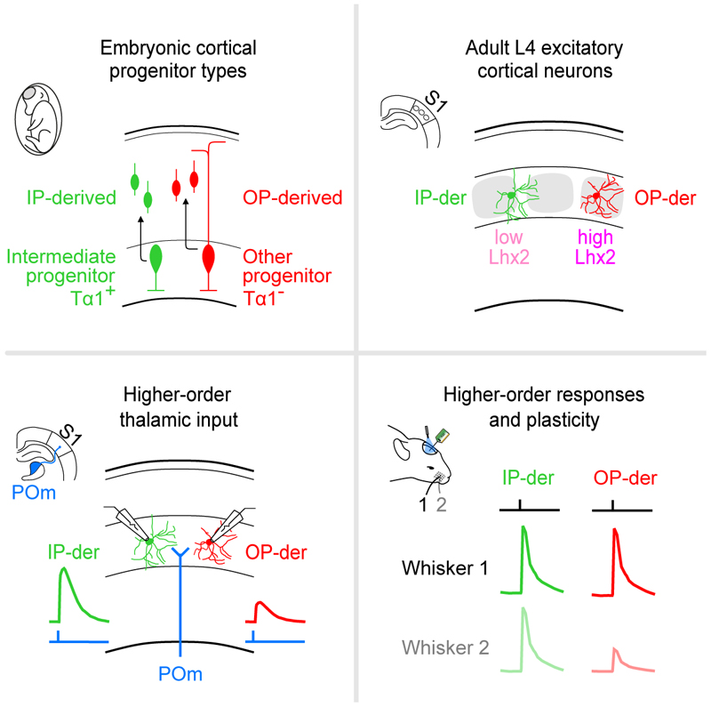
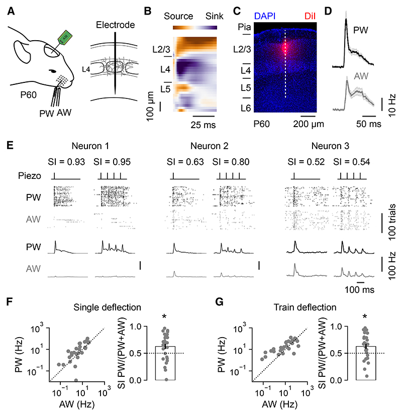
The sensory cortex receives synaptic inputs from both first-order and higher-order thalamic nuclei. First-order inputs relay simple stimulus properties from the periphery, whereas higher-order inputs relay more complex response properties, provide contextual feedback, and modulate plasticity. Here, we reveal that a cortical neuron's higher-order input is determined by the type of progenitor from which it is derived during embryonic development. Within layer 4 (L4) of the mouse primary somatosensory cortex, neurons derived from intermediate progenitors receive stronger higher-order thalamic input and exhibit greater higher-order sensory responses. These effects result from differences in dendritic morphology and levels of the transcription factor Lhx2, which are specified by the L4 neuron's progenitor type. When this mechanism is disrupted, cortical circuits exhibit altered higher-order responses and sensory-evoked plasticity. Therefore, by following distinct trajectories, progenitor types generate diversity in thalamocortical circuitry and may provide a general mechanism for differentially routing information through the cortex.

Keywords: CP: Neuroscience; cortex; development; higher-order; in utero labelling; intermediate progenitor; lineage; optotagging; sensory-evoked plasticity; thalamus.


Conflict of interest statement

Declaration of interests The authors declare no competing interests.

Figures

Graphical abstract
Graphical abstract
Figure 1
Figure 1. S1 L4 neurons differ in their multi-whisker response properties
(A)The activity of individual regular-spiking L4 neurons in S1 was recorded in response to deflection of the PW or AW at P60. (B) L4 was identified using the current source density profile following PW deflection. (C) Histology confirmed the recording location in S1. The dashed line indicates electrode penetration. (D) Mean L4 neuronal responses from an individual animal following a single deflection of the PW (top) or AW (bottom). Shading indicates SEM around the mean. (E) Three L4 neurons with different SI, as defined by relative response to the PW and AW. Raster plots and corresponding peri-stimulus time histograms (PSTHs) show spiking activity over 100 trials of either a single deflection (inner left) or train deflection (inner right) of the PW (black) or AW (gray). (F) Responses of individual neurons to single deflection of the PW and AW (left) and the distribution of corresponding SI values (right). L4 neurons were selective for the PW (n = 28 neurons, p = 0.006, one-sample t test). (G) Responses of individual neurons to train deflection of the PW and AW (left) and the corresponding SI values (right). L4 neurons were selective for the PW (n = 28 neurons, p = 0.006, one-sample t test). Data are represented as mean ± SEM. Scale bars are indicated.
Figure 2
Figure 2. IP-derived L4 neurons exhibit greater multi-whisker responses
(A) IUE of a Tα1-Cre and floxed ChR2-YFP plasmid was used to optotag IP-derived L4 neurons in S1. (B) IP-derived S1 L4 neurons expressing ChR2-YFP at P60. (C) The spiking activity of optotagged IP-derived L4 neurons and neighboring (non-optotagged) unla-beled L4 neurons was recorded in response to the deflection of the PW or AW. (D) Mean responses from an individual animal following a single deflection of the PW (left) or AW (right). (E) Spiking of an individual IP-derived (left, green) and an unlabeled L4 neuron (right, black) over 100 trials of either single deflection (inner left) or train deflection (inner right) of the PW or AW. (F) Responses of individual IP-derived and unlabeled L4 neurons to single deflection of the PW and AW (left) and the distribution of corresponding SI values (right). IP-derived L4 neurons were less selective to the PW, and therefore relatively more responsive to the AW, when compared to unlabeled neurons (n = 29 and 26, p = 0.017, t test). (G) Responses to train deflection of the PW and AW (left) and corresponding SI values (right). IP-derived L4 neurons were less selective to the PW and therefore relatively more responsive to the AW (n = 29 and 26, p = 0.040, t test). Data are represented as mean ± SEM; n = neurons; conventions as in Figure 1. Scale bars are indicated.
Figure 3
Figure 3. IP-derived L4 neurons receive greater input from higher-order thalamus
(A) Experimental design for studying thalamic input to progenitor type-defined L4 neurons. IUE of a Tα1-Cre and two-color Cre-dependent reporter plasmid was used to label IP-derived (GFP expressing, green) and OP-derived (tdTo-mato expressing, red) L4 neurons in S1 (left). At P21, the mice received a thalamic injection of an AAV encoding CAG-ChR2-GFP into either VPM or POm (right). (B) Coronal brain slice through the thalamus at P60 in a VPM-injected animal, with a corresponding section from a brain atlas overlaid (bottom). Histological analysis revealed that 77.2% ± 3.5% (n = 6) of ChR2-GFP expression was restricted to VPM (top). (C) Coronal brain slice through the thalamus in a POm-injected animal, with a corresponding section from brain atlas overlaid (bottom). Histological analysis revealed that 80.4% ± 2.5% (n = 8) of ChR2-GFP expression was restricted to POm (top). (D) To measure VPM input, simultaneous whole-cell recordings were performed from neuronal pairs comprising an IP-derived and an OP-derived L4 neuron in acute slices, while ChR2-GFP-expressing VPM axons were stimulated with light pulses. (E) EPSP peak amplitudes for pairs of IP-derived and OP-derived neurons in response to light stimulation of VPM axons. (F) IP-derived neurons received weaker VPM input than OP-derived neurons (n = 18, p < 0.001, one-sample t test). (G) A similar arrangement was used to measure POm input. (H) EPSP peak amplitudes for pairs of IP-derived and OP-derived neurons in response to light stimulation of POm axons. (I) POm input was biased toward IP-derived neurons, which received stronger POm input than OP-derived neurons (n = 21, p = 0.002, one-sample t test). Data are represented as mean ± SEM; n = animals or neuron pairs. Scale bars are indicated.
Figure 4
Figure 4. Progenitor type predicts differences in how L4 dendritic morphology relates to barrels
(A) Thalamic axonal input to S1 from VPM (left) and POm (right), visualized with ChR2-GFP. (B) IUE of a Tα1-Cre and two-color Cre-dependent reporter plasmid was used to label IP-derived (green) and OP-derived (red) L4 neurons in S1. (C) VGLUT2 immunohistochemistry at P21 (left) was used to relate the distribution of labeled soma to the organization of barrels and septa. A soma position index was defined, with a value of 1 indicating a soma in the center of a barrel (right). Dashed lines indicate outlines of barrels. (D) There was no difference in the distribution of IP-derived or OP-derived L4 neurons (n = 537 and 351, p = 0.827, Mann-Whitney U test). (E) Biocytin fills were used to reconstruct the dendritic morphology of IP-derived and OP-derived neurons. (F) Example image and corresponding reconstruction of an IP-derived (left) and an OP-derived (right) L4 neuron. (G) Dendrites of OP-derived neurons were more likely to target the principal barrel than IP-derived neurons (left; n = 17 and 16, p = 0.005, Mann-Whitney U test). Dendrites of both populations exhibited a similar degree of overlap with the adjacent barrel (center; n = 17 and 16, p = 0.145, Mann-Whitney U test). Dendrites of IP-derived neurons were more likely to target the area outside of barrels, including septa (right; n = 17 and 16, p = 0.004, Mann-Whitney U test). Data are represented as mean ± SEM; n = neurons. Scale bars are indicated.
Figure 5
Figure 5. Progenitor type determines higher-order thalamic input via neuronal Lhx2 levels
(A) IP-derived (green) and OP-derived (red) L4 neurons were labeled in S1, and quantitative immunohistochemistry (IHC) for Lhx2 was performed at P7. (B) IP-derived neurons expressed lower levels of Lhx2 compared to neighboring OP-derived neurons. (C) Lhx2 expression was relatively low in IP-derived neurons compared to neighboring OP-derived neurons (right; n = 88, p < 0.001, one-sample t test). (D) IUE labeling of IP-derived and OP-derived L4 neurons was combined with a CAG-Lhx2 plasmid to increase Lhx2 expression levels. (E) Biocytin fills at P21 were used to reconstruct the dendritic morphology of IP-derived L4 neurons overexpressing Lhx2. (F) Example image and corresponding reconstruction of an IP-derived Lhx2 neuron. (G) Dendrites of IP-derived Lhx2 neurons were more likely to target the principal barrel compared to WT (left; WT data from Figure 3; n = 17 and 15, p = 0.006, Mann-Whitney U test). Dendrites of both populations exhibited a similar degree of overlap with the adjacent barrel (center; n = 17 and 15, p = 0.727, Mann-Whitney U test). Dendrites of IP-derived Lhx2 neurons were less likely to target the area outside of barrels, including septa, compared to WT (right; n = 17 and 15, p = 0.004, Mann-Whitney U test). (H) To study thalamic inputs, IUE was performed as in (D), then mice received a thalamic injection at P21 of an AAV encoding CAG-ChR2-GFP into either VPM or POm. (I) EPSP peak amplitudes for pairs of IP-derived and OP-derived Lhx2-overexpressing neurons in response to light stimulation of VPM axons. (J) No significant bias was detected in the strength of VPM input (n = 16, p = 0.06, one-sample t test). (K) EPSP peak amplitudes for pairs of IP-derived and OP-derived Lhx2 neurons in response to light stimulation of POm axons. (L) No significant bias was detected in the strength of POm input (n = 22, p = 0.687, one-sample t test). Data are represented as mean ± SEM; n = neurons/neuron pairs. Scale bars are indicated.
Figure 6
Figure 6. Progenitor-specified higher-order circuits contribute to multi-whisker responsivity
(A) IUE of a Tα1-Cre, floxed ChR2-YFP, and CAG-Lhx2 plasmid was used to optotag IP-derived Lhx2 L4 neurons in S1. (B) ChR2-YFP expressing IP-derived Lhx2 L4 neurons at P60. (C) The spiking activity of optotagged IP-derived Lhx2 L4 neurons was recorded in response to the deflection of the PW or AW. (D) Mean responses of IP-derived WT L4 neurons and IP-derived Lhx2 L4 neurons. (E) Spiking of an individual IP-derived WT neuron (left) and Lhx2 neuron (right) over 100 trials of either a single deflection (inner left) or trains of deflection (inner right) of the PW or AW. (F) Responses of individual IP-derived WT and Lhx2 L4 neurons to single deflection of the PW and AW (left) and the distribution of corresponding SI values (right). Compared to the WT, IP-derived Lhx2 L4 neurons showed greater selectivity for the PW and less responsivity to the AW (n = 29, 19; p = 0.011, Mann Whitney U test). (G) Responses to train deflection of the PW and AW (left) and corresponding SI values (right). Compared to WT, IP-derived Lhx2 L4 neurons showed greater selectivity for the PW and less responsivity to the AW (n = 29, 19; p = 0.014, t test). Data are represented as mean ± SEM; n = neurons; conventions as in Figure 2. Scale bars are indicated.
Figure 7
Figure 7. Progenitor-specified higher-order circuits support sensory-evoked plasticity
(A) IP-derived WT L4 neurons underwent IUE of a Tα1-Cre and floxed ChR2-YFP plasmid. IP-derived Lhx2 L4 neurons underwent IUE of the same plasmids plus a CAG-Lhx2 plasmid. (B) Sensory-evoked plasticity was examined in L2/3 of S1 at P28 using a rhythmic whisker stimulation (RWS) protocol (8 Hz for 60 s). The ChR2-YFP expression enabled us to use the response to a light pulse to confirm that the recording was targeting a region of S1 containing electroporated L4 neurons. (C) Raster plots and PSTHs show multi-unit L2/3 spiking activity in a WT animal. Responses to whisker deflections (0.1 Hz) are shown before (Pre) and after (Post) RWS. (D) Averaged (left) and separate (right) population data from WT animals reveal that RWS potentiated L2/3 activity (n = 6, p = 0.007, paired t test). (E) Multi-unit L2/3 activity in an Lhx2 animal. (F) Lhx2 animals did not exhibit potentiation of L2/3 activity following RWS (n = 7, p = 0.548, paired t test). (G) Normalized L2/3 multi-unit activity relative to the time of RWS (each data point is the mean of five whisker deflections, 0.1 Hz). Shading indicates SEM. (H) Delta spike rate in WT control animals that did not experience the RWS protocol (Con), WT animals that experienced RWS (WT+RWS), or Lhx2 animals that experienced RWS (Lhx2+RWS) (n = 4, 6, and 7; p = 0.001, Kruskal-Wallis test; WT control vs. WT+RWS, p < 0.05; WT+RWS vs. Lhx2+RWS, p < 0.05, WT control vs. Lhx2+RWS, p > 0.05, Dunn’s test). Data are represented as mean ± SEM; n = animals.

Similar articles

Cited by

  • Sensory modality-specific wiring of thalamocortical circuits.
    Guillamón-Vivancos T, Aníbal-Martínez M, Puche-Aroca L, Martini FJ, López-Bendito G. Guillamón-Vivancos T, et al. Nat Rev Neurosci. 2025 Jul 30. doi: 10.1038/s41583-025-00945-y. Online ahead of print. Nat Rev Neurosci. 2025. PMID: 40745219 Review.

References

    1. Bruno RM, Simons DJ. Feedforward Mechanisms of Excitatory and Inhibitory Cortical Receptive Fields. J Neurosci. 2002;22:10966–10975. doi: 10.1523/JNEUROSCI.22-24-10966.2002. - DOI - PMC - PubMed
    1. Hubel DH, Wiesel TN. Receptive fields, binocular interaction and functional architecture in the cat’s visual cortex. J Physiol. 1962;160:106–154. doi: 10.1113/jphysiol.1962.sp006837. - DOI - PMC - PubMed
    1. Reid RC, Alonso JM. Specificity of monosynaptic connections from thalamus to visual cortex. Nature. 1995;378:281–284. - PubMed
    1. Kwegyir-Afful EE, Bruno RM, Simons DJ, Keller A. The Role of Thalamic Inputs in Surround Receptive Fields of Barrel Neurons. J Neurosci. 2005;25:5926–5934. doi: 10.1523/JNEUROSCI.1360-05.2005. - DOI - PMC - PubMed
    1. Sherman SM, Guillery RW. On the actions that one nerve cell can have on another: Distinguishing “drivers” from “modulators”. Proc Natl Acad Sci USA. 1998;95:7121–7126. doi: 10.1073/pnas.95.12.7121. - DOI - PMC - PubMed

Publication types

LinkOut - more resources