. 2024 Jun:327:114943.
doi: 10.1016/j.jviromet.2024.114943. Epub 2024 Apr 26.

Establishment of Swine Primary Nasal, Tracheal, and Bronchial Epithelial Cell Culture Models for the Study of Influenza Virus Infection

Affiliations

Establishment of Swine Primary Nasal, Tracheal, and Bronchial Epithelial Cell Culture Models for the Study of Influenza Virus Infection

Madelyn Krunkosky et al. J Virol Methods. 2024 Jun.

Abstract

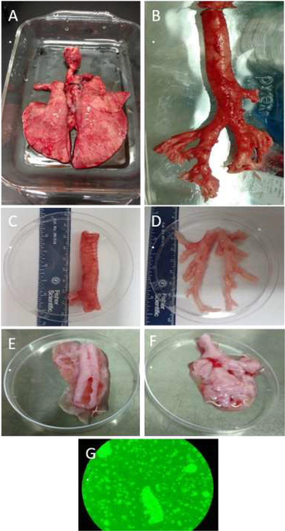
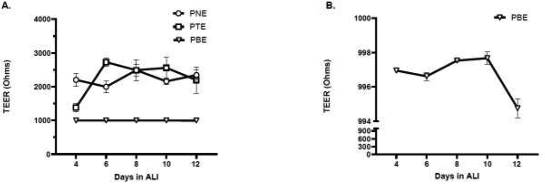
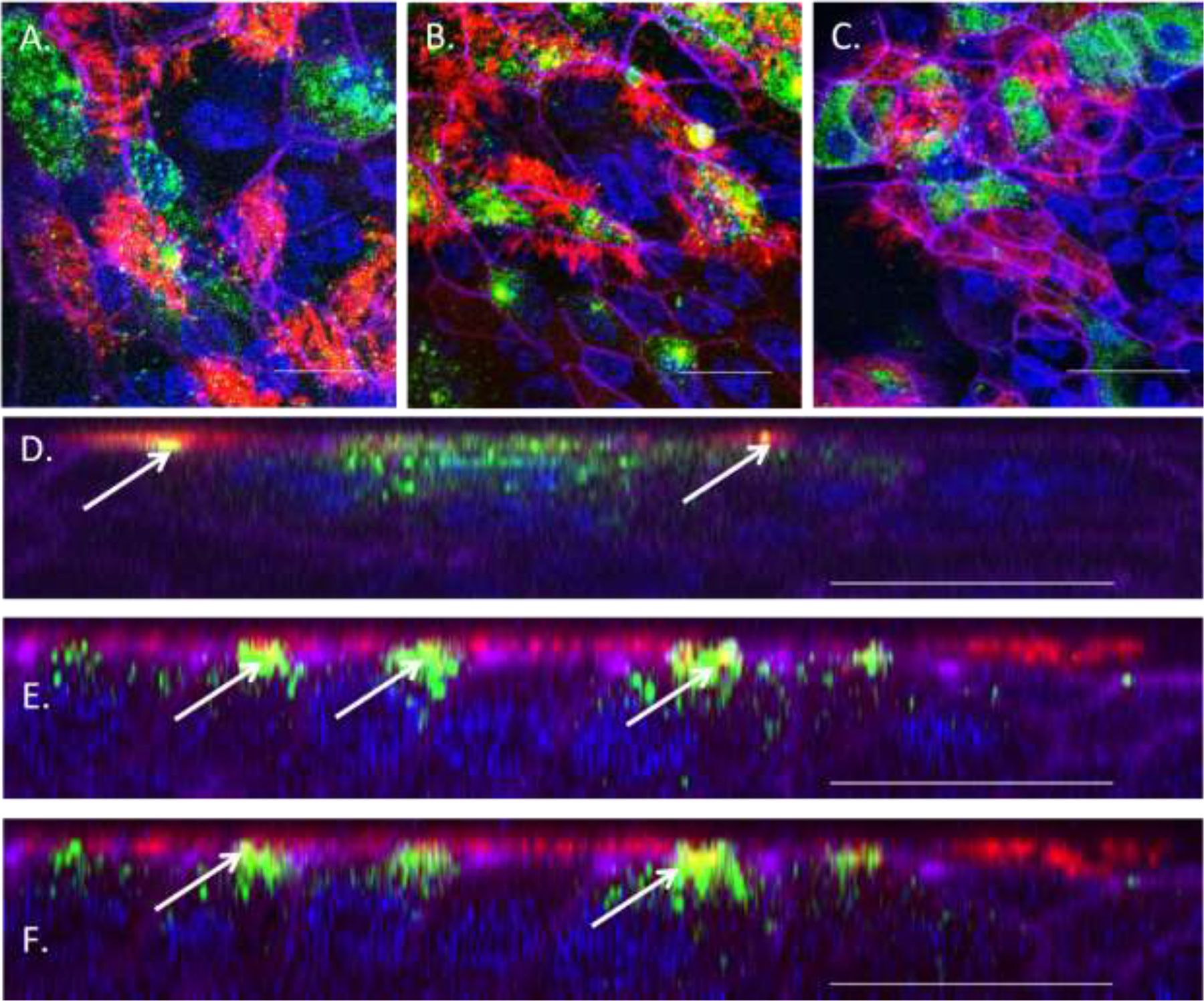
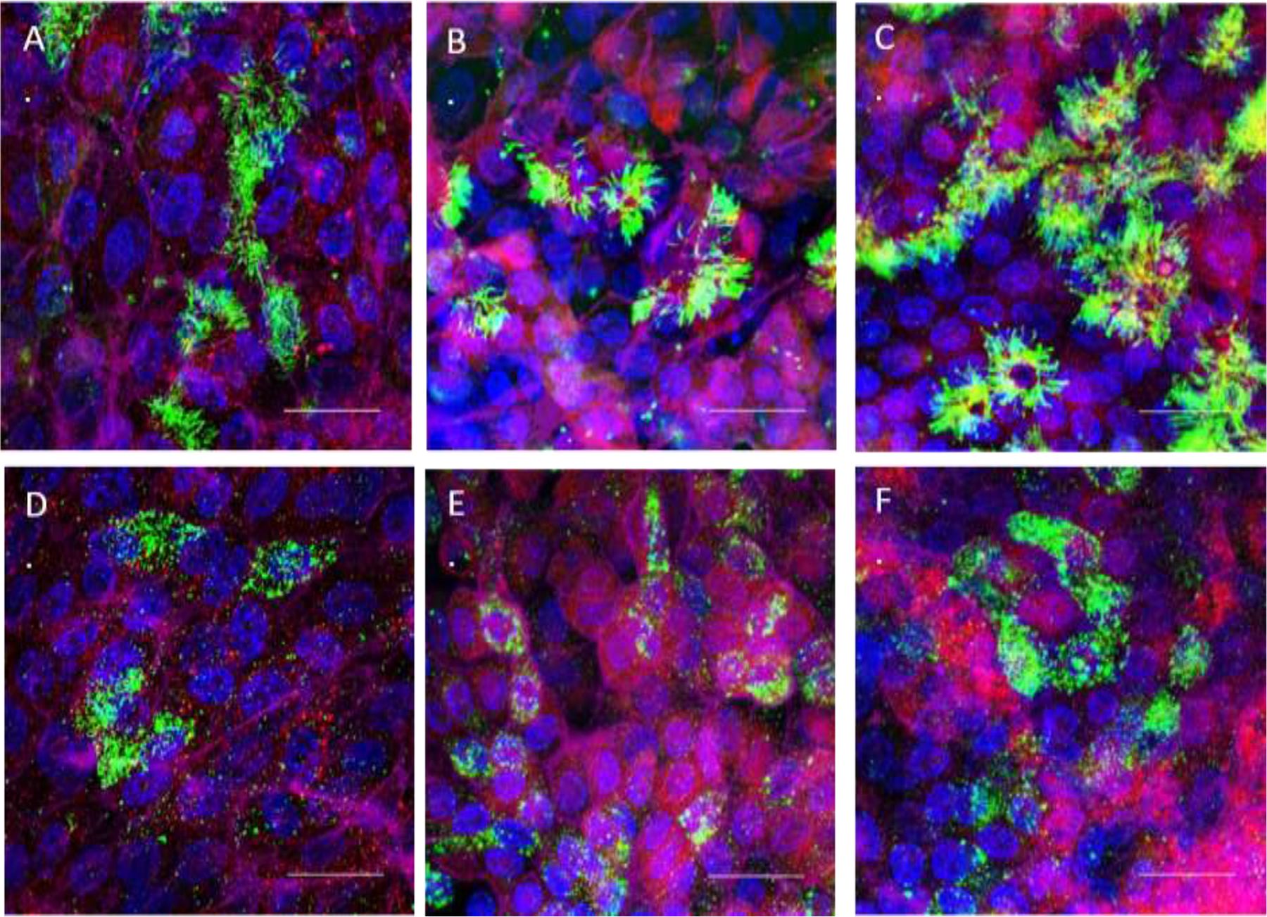
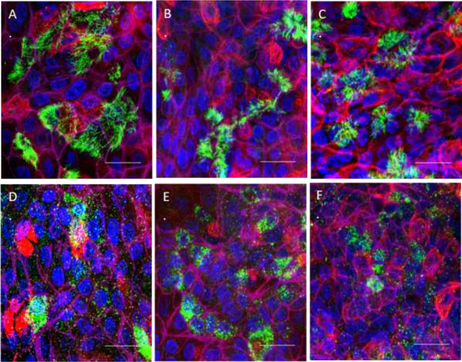
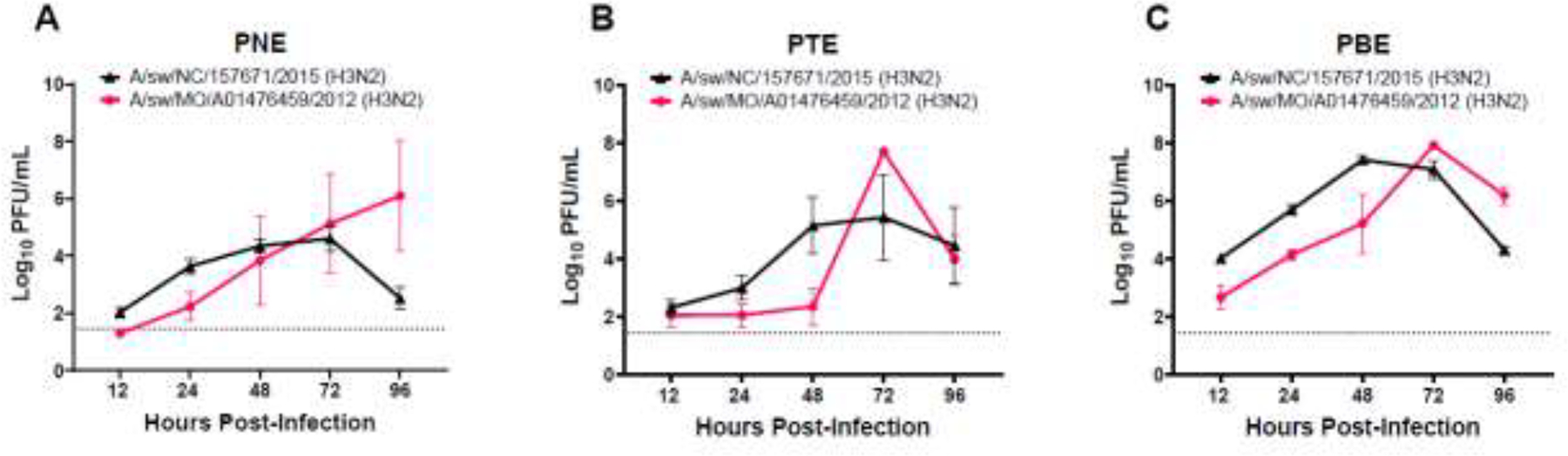
We established primary porcine nasal, tracheal, and bronchial epithelial cells that recapitulate the physical and functional properties of the respiratory tract and have the ability to fully differentiate. Trans-well cultures demonstrated increased transepithelial electrical resistance over time the presence of tight junctions as demonstrated by immunohistochemistry. The nasal, tracheal, and bronchial epithelial cells developed cilia, secreted mucus, and expressed sialic acids on surface glycoproteins, the latter which are required for influenza A virus infection. Swine influenza viruses were shown to replicate efficiently in the primary epithelial cell cultures, supporting the use of these culture models to assess swine influenza and other virus infection. Primary porcine nasal, tracheal, and bronchial epithelial cell culture models enable assessment of emerging and novel influenza viruses for pandemic potential as well as mechanistic studies to understand mechanisms of infection, reassortment, and generation of novel virus. As swine are susceptible to infection with multiple viral and bacterial respiratory pathogens, these primary airway cell models may enable study of the cellular response to infection by pathogens associated with Porcine Respiratory Disease Complex.

Keywords: Epithelial cells; Influenza A virus; Models; N-Acetylneuraminic acid; Swine.


Conflict of interest statement

Declaration of Competing Interest The authors declare that they have no known competing financial interests or personal relationships that could have appeared to influence the work reported in this paper.

Figures

Figure 1:
Figure 1:. Harvest and Culture of PTE and PBE Cells.
A. Pluck harvested from a 4–6-week-old pig B. Dissected out trachea and bronchi intact C. Dissected trachea D. Dissected bronchi and bronchioles E. Post digestion trachea F. Post digestion bronchi and bronchioles G. Single cell suspension with multiple ciliated epithelial rafts 20X objective phase contrast on inverted microscope
Figure 2:
Figure 2:. PNE, PTE, and PBE Trans-epithelial Electrical Resistance.
A. Trans-epithelial electrical resistance (TEERs) of differentiated swine cells cultured at air-liquid interface (ALI). B. TEER values for PBE cells, with y axis expanded to highlight changes at day 12. Data represents one independent experiment. Three measurements were taken from each well at each individual time point and averaged. n=6 wells per time-point +/− SEM.
Figure 3:
Figure 3:. PNE, PTE, and PBE Immunohistochemistry of Tight Junctions.
Claudin 4 and ZO-1 to detect presence of tight junctions on primary swine epithelial cells in air-liquid interface (ALI)=green, acetylated tubulin to detect the presence of cilia on primary swine epithelial cells in ALI=red, and Phalloidin to detect colocalization of Claudin 4 and ZO-1 with F-actin=purple, white bar indicates a measurement of 25 microns A. PNE-Claudin 4 and cilia (top view) B. PTE-Claudin 4 and cilia (top view) C. PBE-Claudin 4 and cilia (top view) D. PNE-ZO-1 and cilia E. PTE-ZO-1 and cilia, and F. PBE-ZO-1 and cilia. Figures D-F green represents ZO-1 creating tight junctions on apical surface colocalizing with F-actin. Yellow represents colocalization indicating tight junction formation of F-actin, ZO-1, and cilia basal bodies, which is indicated by the white arrow.
Figure 4:
Figure 4:. α2,6-Sialic Acid Lectin, Cilia, and Mucin Immunohistochemistry of PNE, PTE, and PBE Cells.
SNA lectin to detect presence of α2,6-linked sialic acid receptors on primary swine epithelial cells in air-liquid interface (ALI)=red, beta and acetylated tubulin to detect the presence of cilia on primary swine epithelial cells in ALI=green (A, B, C), Muc5AC to detect the presence of mucus and mucin granules in goblet cells in primary swine epithelial cells in ALI=green (D, E, F), DAPI, nuclear stain=blue, and Phalloidin to detect the presence of tight junctions, F-actin=purple, white bar indicates a measurement of 25 microns A. PNE-SNA and cilia B. PTE-SNA and cilia C. PBE-SNA and cilia D. PNE-SNA and mucus E. PTE-SNA and mucus, and F. PBE SNA and mucus.
Figure 5:
Figure 5:. α2,3-Sialic Acid Lectin, Cilia, and Mucin Immunohistochemistry of PNE, PTE, and PBE Cells.
MAL-2 lectin to detect presence of α2,3-linked sialic acid receptors on primary swine epithelial cells in air-liquid interface (ALI)=red, beta and acetylated tubulin to detect the presence of cilia on primary swine epithelial cells in ALI=green (A, B, C), Muc5AC to detect the presence of mucus and mucin granules in goblet cells in primary swine epithelial cells in ALI=green (D, E, F), dapi, nuclear stain=blue, and Phallodin to detect the presence of tight junctions, F-actin=purple, white bar indicates a measurement of 25 microns A. PNE-MAL-2 and cilia B. PTE-MAL-2 and cilia C. PBE-MAL-2 and cilia D. PNE-MAL-2 and mucus E. PTE-MAL-2 and mucus, and F. PBE MAL-2 and mucus.
Figure 6:
Figure 6:. Replication Kinetics of Swine Isolates Containing Gene Segments of Swine and Human Origin in PNE, PTE, and PBE Cells.
PNE, PTE, and PBE cells were grown on transwells and allowed to fully differentiate in ALI. Once cilia and mucus secretion were present, cells were infected with A/sw/NC/157671/2015 (H3N2) or A/sw/MO/A01476459/2012 (H3N2) at 250 PFU/well A. PNE B. PTE, and C. PBE. Data represents one independent experiment. For each virus the same set of wells was used for all subsequent time-points. Error bars represent +/− SEM of triplicate wells.

Similar articles

Cited by

References

    1. Brockmeier SL, Halbur PG, Thacker EL, 2002. Porcine Respiratory Disease Complex Chapter 13. Polymicrobial Diseases.
    1. Byrd-Leotis L, Cummings RD and Steinhauer DA, 2017. The Interplay between the Host Receptor and Influenza Virus Hemagglutinin and Neuraminidase. Int J Mol Sci 18, 1541. - PMC - PubMed
    1. Chan RW, Karamanska R, Van Poucke S, Van Reeth K, Chan IW, Chan MC, Dell A, Peiris JS, Haslam SM, Guan Y and Nicholls JM, 2013. Infection of swine ex vivo tissues with avian viruses including H7N9 and correlation with glycomic analysis. Influenza Other Respir Viruses 7, 1269–82. - PMC - PubMed
    1. Curran SJ, Griffin EF, Ferreri LM, Kyriakis CS, Howerth EW, Perez DR and Tompkins SM, 2024. Swine influenza A virus isolates containing the pandemic H1N1 origin matrix gene elicit greater disease in the murine model. Microbiol Spectr, e0338623. - PMC - PubMed
    1. Fulcher ML, Gabriel S, Burns KA, Yankaskas JR and Randell SH, 2005. Well-differentiated human airway epithelial cell cultures. Methods Mol Med 107, 183–206. - PubMed

Publication types

LinkOut - more resources