

RNA viromes of Dermacentor nuttalli ticks reveal a novel uukuvirus in Qīnghăi Province, China
- PMID: 38679334
- PMCID: PMC11401450
- DOI: 10.1016/j.virs.2024.04.006
RNA viromes of Dermacentor nuttalli ticks reveal a novel uukuvirus in Qīnghăi Province, China
Abstract
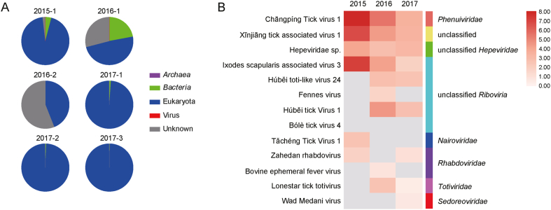
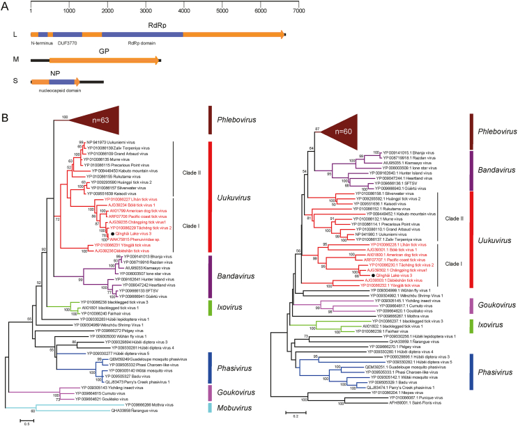
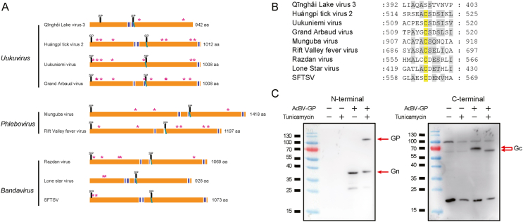
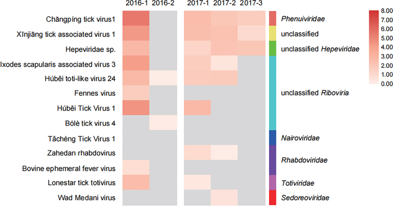
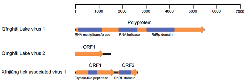
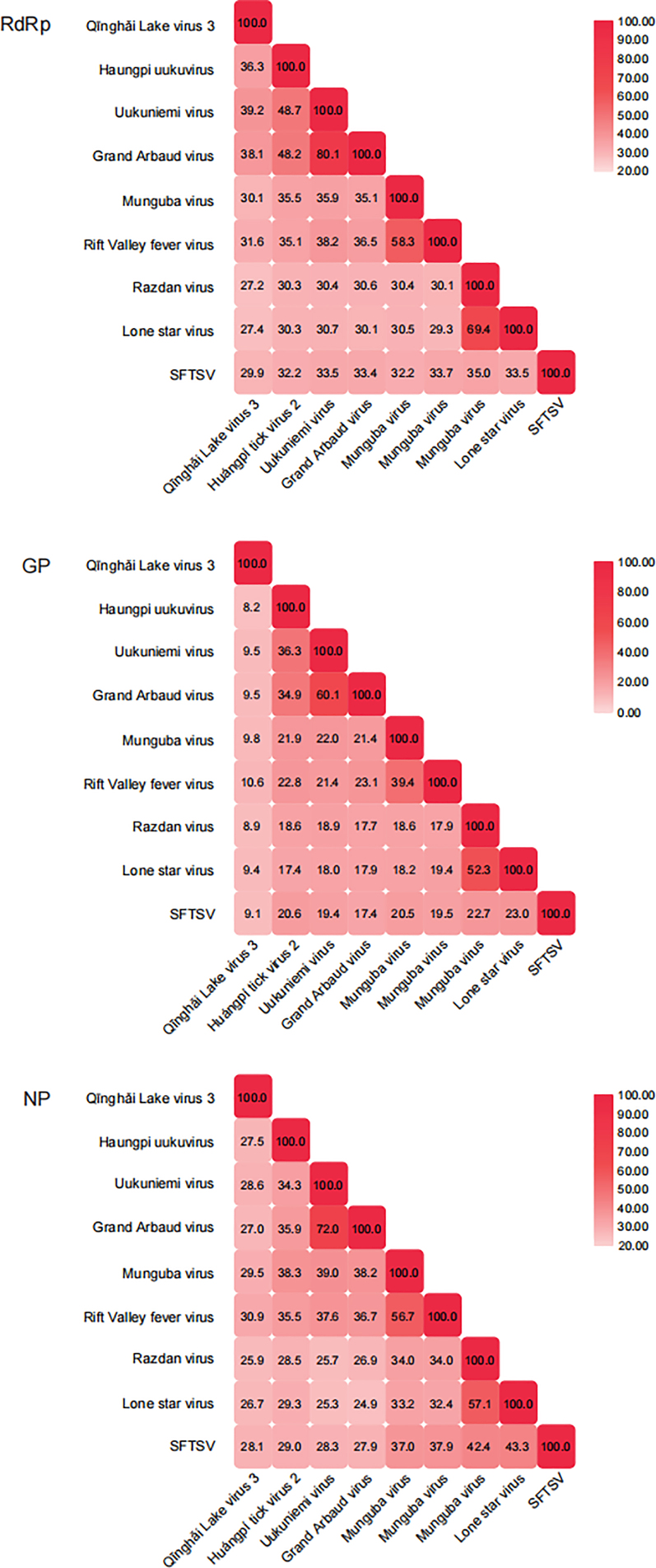
Ticks are a major parasite on the Qīnghăi-Tibet Plateau, western China, and represent an economic burden to agriculture and animal husbandry. Despite research on tick-borne pathogens that threaten humans and animals, the viromes of dominant tick species in this area remain unknown. In this study, we collected Dermacentor nuttalli ticks near Qīnghăi Lake and identified 13 viruses belonging to at least six families through metagenomic sequencing. Four viruses were of high abundance in pools, including Xīnjiāng tick-associated virus 1 (XJTAV1), and three novel viruses: Qīnghăi Lake virus 1, Qīnghăi Lake virus 2 (QHLV1, and QHLV2, unclassified), and Qīnghăi Lake virus 3 (QHLV3, genus Uukuvirus of family Phenuiviridae in order Bunyavirales), which lacks the M segment. The minimum infection rates of the four viruses in the tick groups were 8.2%, 49.5%, 6.2%, and 24.7%, respectively, suggesting the prevalence of these viruses in D. nuttalli ticks. A putative M segment of QHLV3 was identified from the next-generation sequencing data and further characterized for its signal peptide cleavage site, N-glycosylation, and transmembrane region. Furthermore, we probed the L, M, and S segments of other viruses from sequencing data of other tick pools by using the putative M segment sequence of QHLV3. By revealing the viromes of D. nuttalli ticks, this study enhances our understanding of tick-borne viral communities in highland regions. The putative M segment identified in a novel uukuvirus suggests that previously identified uukuviruses without M segments should have had the same genome organization as typical bunyaviruses. These findings will facilitate virus discovery and our understanding of the phylogeny of tick-borne uukuviruses.
Keywords: Dermacentor nuttalli; M segment; Qīnghăi Province; Uukuvirus; Virome.
Copyright © 2024 The Authors. Publishing services by Elsevier B.V. All rights reserved.
Conflict of interest statement
Conflict of interest Prof. Fei Deng is an editorial board member for Virologica Sinica and was not involved in the editorial review or the decision to publish this article. The authors declare that they have no conflict of interest.
Figures
References
-
- Boulanger N., Boyer P., Talagrand-Reboul E., Hansmann Y. Ticks and tick-borne diseases. Med. Maladies Infect. 2019;49:87–97. - PubMed
-
- Chitimia L., Lin R.Q., Cosoroaba I., Braila P., Song H.Q., Zhu X.Q. Molecular characterization of hard and soft ticks from Romania by sequences of the internal transcribed spacers of ribosomal DNA. Parasitol. Res. 2009;105:907–911. - PubMed
MeSH terms
Substances
LinkOut - more resources
Full Text Sources
