

Giant intraperitoneal non-pancreatic pseudocyst: a case report
- PMID: 38679699
- PMCID: PMC11057145
- DOI: 10.1186/s13256-024-04503-5
Giant intraperitoneal non-pancreatic pseudocyst: a case report
Abstract
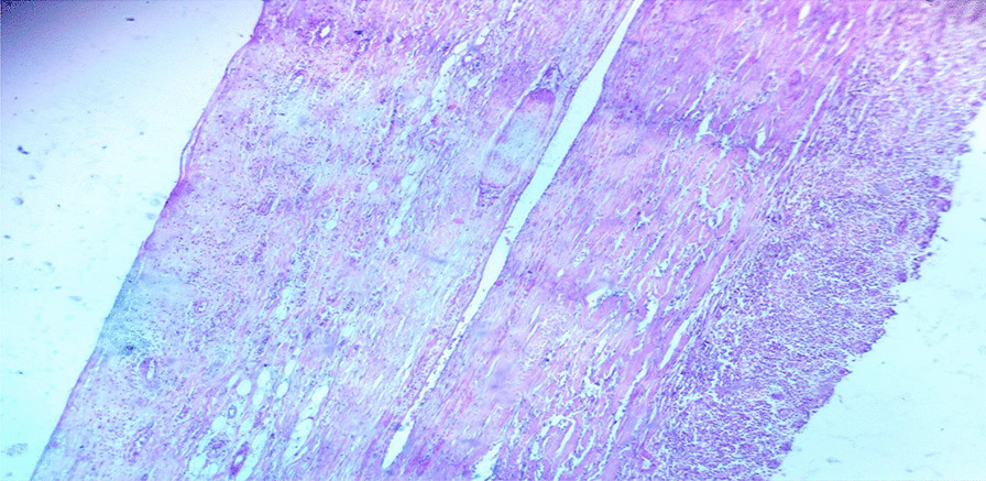
Introduction: Non-pancreatic pseudocysts are rare lesions that typically form from the omentum and mesentery. These cysts have a thick fibrotic wall made up of fibrous tissue and may show signs of calcifications and inflammatory changes. The fluid inside them can vary, ranging from hemorrhage and pus to serous or sometimes chylous content. In most cases, these cysts appear as a result of trauma, surgery, or infection.
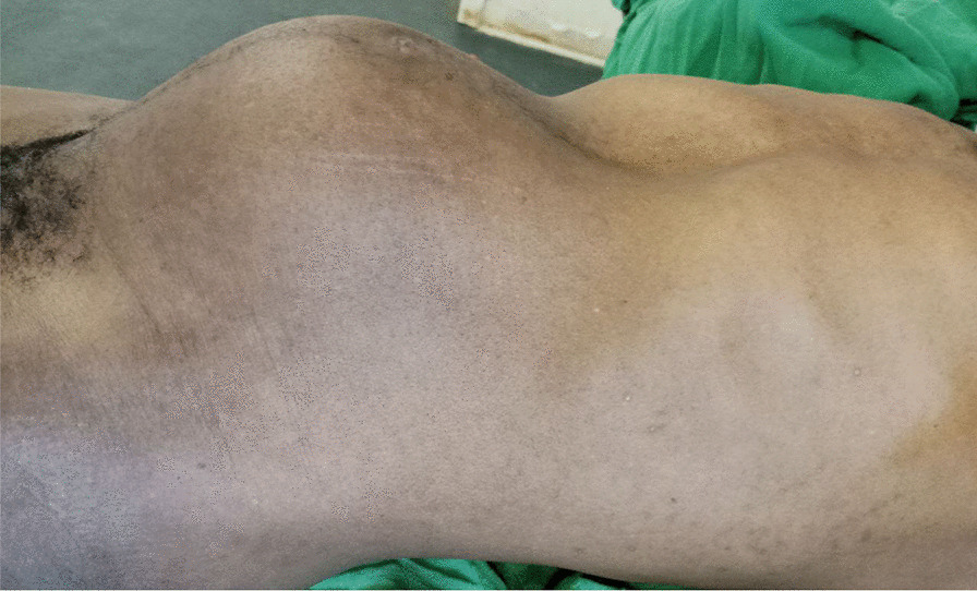
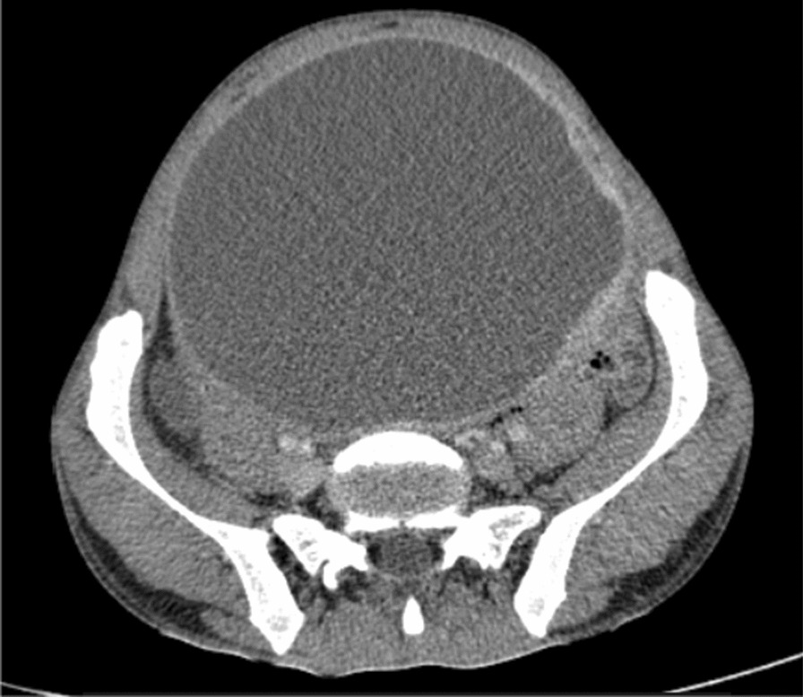
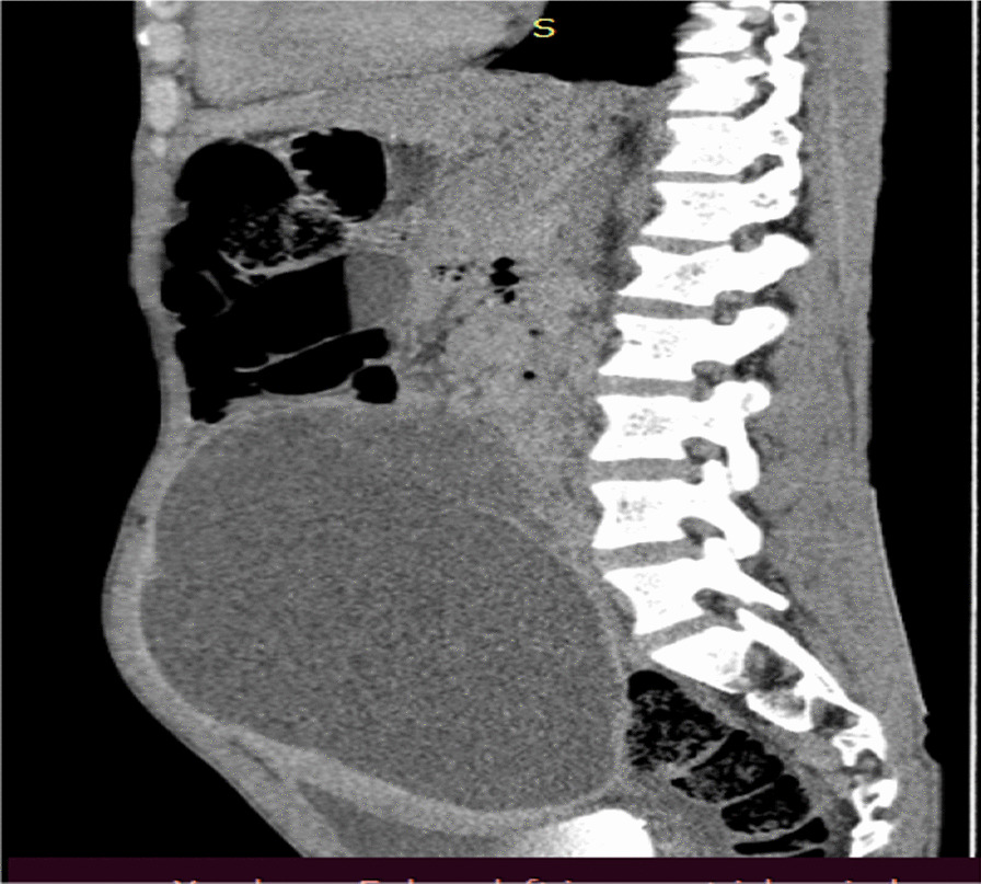
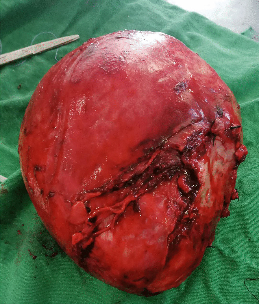
Case presentation: A 35-year-old male patient from Ethiopia presented with swelling in his lower abdomen that had been present for 2 years. Initially, the swelling was small but gradually increased in size. The patient experienced frequent urination but no pain or difficulty during urination, urgency, intermittent urination, or blood in the urine. The swelling was initially painless but became painful 2 months prior to his presentation. Abdominal computed tomography scans revealed a well-defined, lobulated peritoneal lesion measuring 16 × 12 × 10 cm, consisting primarily of fluid-filled cysts with a thick, enhancing wall and septa. Additionally, there was a large, heterogeneous enhancing soft tissue component measuring 8 × 6 cm. As a result, the cystic mass was surgically removed in its entirety with partial removal of the bladder wall, and the patient was discharged in an improved condition.
Conclusion: Primary non-pancreatic pseudocysts are extremely rare lesions that must be differentiated from other possible causes of cystic lesions within the peritoneal or retroperitoneal regions. Surgeons should be aware of the potential occurrence of these lesions, which may have an unknown origin.
Keywords: Bladder; Case report; Inflammatory; Non-pancreatic; Pseudocyst.
© 2024. The Author(s).
Conflict of interest statement
No potential conflict of interest relevant to this article was reported.
Figures


References
-
- Chawla VD, Saraswat A, Tauheed F, Gupta PD, Chauhan VS. Giant benign retroperitoneal non-pancreatic pseudocyst in a female: a case report & a diagnostic challenge. Eur J Mol Clin Med. 2021;8(04):2021.
Publication types
MeSH terms
LinkOut - more resources
Full Text Sources
Medical
Research Materials
