

Mass Spectrometry Reveals Molecular Effects of Citrulline Supplementation during Bone Fracture Healing in a Rat Model
- PMID: 38679918
- PMCID: PMC11157653
- DOI: 10.1021/jasms.4c00028
Mass Spectrometry Reveals Molecular Effects of Citrulline Supplementation during Bone Fracture Healing in a Rat Model
Abstract
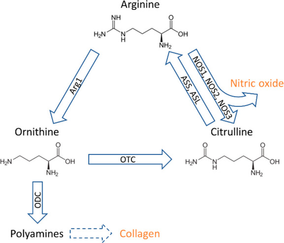
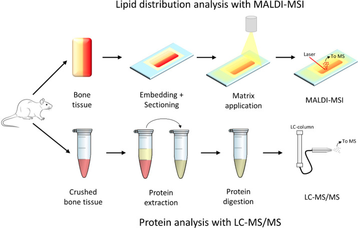
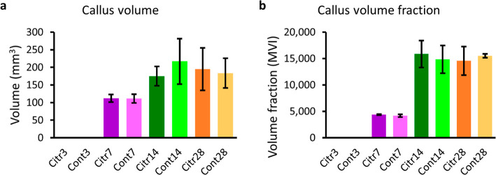
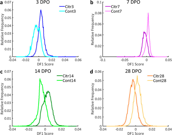
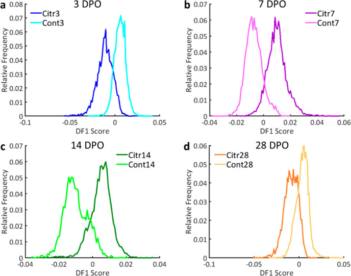
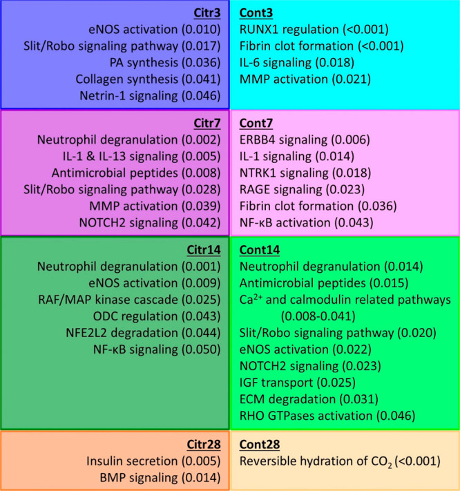
Bone fracture healing is a complex process in which specific molecular knowledge is still lacking. The citrulline-arginine-nitric oxide metabolism is one of the involved pathways, and its enrichment via citrulline supplementation can enhance fracture healing. This study investigated the molecular effects of citrulline supplementation during the different fracture healing phases in a rat model. Microcomputed tomography (μCT) was applied for the analysis of the fracture callus formation. Matrix-assisted laser desorption/ionization mass spectrometry imaging (MALDI-MSI) and liquid-chromatography tandem mass spectrometry (LC-MS/MS) were used for lipid and protein analyses, respectively. μCT analysis showed no significant differences in the fracture callus volume and volume fraction between the citrulline supplementation and control group. The observed lipid profiles for the citrulline supplementation and control group were distinct for the different fracture healing stages. The main contributing lipid classes were phosphatidylcholines (PCs) and lysophosphatidylcholines (LPCs). The changing effect of citrulline supplementation throughout fracture healing was indicated by changes in the differentially expressed proteins between the groups. Pathway analysis showed an enhancement of fracture healing in the citrulline supplementation group in comparison to the control group via improved angiogenesis and earlier formation of the soft and hard callus. This study showed the molecular effects on lipids, proteins, and pathways associated with citrulline supplementation during bone fracture healing, even though no effect was visible with μCT.
Conflict of interest statement
The authors declare no competing financial interest.
Figures
Similar articles
-
Enhancement of fracture healing after citrulline supplementation in mice.Eur Cell Mater. 2020 Mar 20;39:183-192. doi: 10.22203/eCM.v039a12. Eur Cell Mater. 2020. PMID: 32195554
-
The effects of combined human parathyroid hormone (1-34) and zoledronic acid treatment on fracture healing in osteoporotic rats.Osteoporos Int. 2012 Apr;23(4):1463-74. doi: 10.1007/s00198-011-1751-6. Epub 2011 Sep 3. Osteoporos Int. 2012. PMID: 21892678
-
Zoledronic acid suppresses callus remodeling but enhances callus strength in an osteoporotic rat model of fracture healing.Bone. 2015 Dec;81:702-711. doi: 10.1016/j.bone.2015.09.018. Epub 2015 Oct 3. Bone. 2015. PMID: 26434668
-
Nano-copper-bearing stainless steel promotes fracture healing by accelerating the callus evolution process.Int J Nanomedicine. 2017 Nov 27;12:8443-8457. doi: 10.2147/IJN.S146866. eCollection 2017. Int J Nanomedicine. 2017. PMID: 29225463 Free PMC article.
-
Fracture healing with alendronate treatment in the Brtl/+ mouse model of osteogenesis imperfecta.Bone. 2013 Sep;56(1):204-12. doi: 10.1016/j.bone.2013.06.003. Epub 2013 Jun 14. Bone. 2013. PMID: 23774443 Free PMC article.
Cited by
-
Molecular Determinants of Bone Plasticity Regeneration After Trauma: Forensic Consequences.Int J Mol Sci. 2025 Jul 25;26(15):7184. doi: 10.3390/ijms26157184. Int J Mol Sci. 2025. PMID: 40806316 Free PMC article. Review.
-
Diagnostic value of LncRNA SNHG16 for osteoporotic fractures and its potential regulation of fracture healing.Hereditas. 2025 Apr 7;162(1):54. doi: 10.1186/s41065-025-00423-6. Hereditas. 2025. PMID: 40197315 Free PMC article.
References
-
- Oryan A.; Monazzah S.; Bigham-Sadegh A. Bone injury and fracture healing biology. Biomed Environ. Sci. 2015, 28 (1), 57–71. - PubMed
-
- Tosounidis T.; Kontakis G.; Nikolaou V.; Papathanassopoulos A.; Giannoudis P. V. Fracture healing and bone repair: an update. Trauma 2009, 11 (3), 145–156. 10.1177/1460408609335922. - DOI
MeSH terms
Substances
LinkOut - more resources
Full Text Sources
