Review
. 2024 Apr 12:15:1395035.
doi: 10.3389/fimmu.2024.1395035. eCollection 2024.

MNDA, a PYHIN factor involved in transcriptional regulation and apoptosis control in leukocytes

Affiliations
Review

MNDA, a PYHIN factor involved in transcriptional regulation and apoptosis control in leukocytes

Stefania Bottardi et al. Front Immunol. .

Abstract

Inflammation control is critical during the innate immune response. Such response is triggered by the detection of molecules originating from pathogens or damaged host cells by pattern-recognition receptors (PRRs). PRRs subsequently initiate intra-cellular signalling through different pathways, resulting in i) the production of inflammatory cytokines, including type I interferon (IFN), and ii) the initiation of a cascade of events that promote both immediate host responses as well as adaptive immune responses. All human PYRIN and HIN-200 domains (PYHIN) protein family members were initially proposed to be PRRs, although this view has been challenged by reports that revealed their impact on other cellular mechanisms. Of relevance here, the human PYHIN factor myeloid nuclear differentiation antigen (MNDA) has recently been shown to directly control the transcription of genes encoding factors that regulate programmed cell death and inflammation. While MNDA is mainly found in the nucleus of leukocytes of both myeloid (neutrophils and monocytes) and lymphoid (B-cell) origin, its subcellular localization has been shown to be modulated in response to genotoxic agents that induce apoptosis and by bacterial constituents, mediators of inflammation. Prior studies have noted the importance of MNDA as a marker for certain forms of lymphoma, and as a clinical prognostic factor for hematopoietic diseases characterized by defective regulation of apoptosis. Abnormal expression of MNDA has also been associated with altered levels of cytokines and other inflammatory mediators. Refining our comprehension of the regulatory mechanisms governing the expression of MNDA and other PYHIN proteins, as well as enhancing our definition of their molecular functions, could significantly influence the management and treatment strategies of numerous human diseases. Here, we review the current state of knowledge regarding PYHIN proteins and their role in innate and adaptive immune responses. Emphasis will be placed on the regulation, function, and relevance of MNDA expression in the control of gene transcription and RNA stability during cell death and inflammation.

Keywords: MNDA; PYHIN factors; apoptosis; genotoxic stress; innate immunity; transcription control.


Conflict of interest statement

The authors declare that the research was conducted in the absence of any commercial or financial relationships that could be construed as a potential conflict of interest.

Figures

Figure 1
Figure 1
Human PYHIN proteins. Schematic diagram of the PYHIN protein structure with amino acid position. PYHIN proteins have a conserved domain architecture comprised of one PYRIN domain (PYD; light blue boxes), one or two hematopoietic IFN-inducible nuclear protein with a 200-amino-acid repeat (HIN-200; light orange boxes) domain/s, and at least one intrinsically disordered region (IDR; dark blue boxes). MNDA IDR consists of a stretch of 115/113 amino acids, spanning from residue 90/93 to residue 205/206. Except for AIM2, which has a predominant cytoplasmic distribution, the other PYHIN members contain either a multipartite nuclear localization signal (NLS) (PYHIN1/IFIX and IFI16) or a putative NLS (MNDA) (yellow boxes). IDR prediction was performed with PSIPRED (http://bioinf.cs.ucl.ac.uk/; 01/2024) and the results are plotted as scatter graphs. The canonical sequences of the PYHIN factors and the identification of their conserved domains were retrieved from the national library of medicine (NCBI) (https://www.ncbi.nlm.nih.gov/) and UniProt (https://www.uniprot.org/) databases. Reference sequences are as follows: MNDA: myeloid cell nuclear differentiation antigen [Homo sapiens]; NCBI Reference Sequence: NP_002423.1; UniProt ID: P41218. AIM2: Interferon-inducible protein AIM2 isoform 1 [Homo sapiens]; NCBI Reference Sequence: NP_004824.1; UniProt ID: O14862. IFI16: gamma-interferon-inducible protein 16 isoform 1 [Homo sapiens]; NCBI Reference Sequence: NP_001193496.1; UniProt ID: Q16666. PYHIN1/IFIX: Pyrin and HIN domain-containing protein 1 isoform alpha 1 [Homo sapiens]; NCBI Reference Sequence: NP_689714.2; UniProt ID: Q6K0P9. X-axis: amino acid number and position; Y-axis: confidence score.
Figure 2
Figure 2
Assembly and activation of the AIM2 inflammasome. Overview of AIM2 inflammasome activation. Cytosolic dsDNA from invading pathogens or mislocated self-dsDNA (pathogen- or danger-associated molecular patterns: PAMPs or DAMPs) directly binds to the pattern recognition receptor (PRR) AIM2. This association, which relieves the intramolecular auto-inhibition imposed by the PYD-HIN-200 interaction, allows the recruitment of apoptosis-associated speck-like protein containing a CARD (ASC) as well as the binding and proximity-induced auto-proteolytic maturation of the pro-caspase-1. This inflammasome activation is likely to occur in the cytoplasmic compartment of the cell, where AIM2 is primarily located. Active caspase-1 can then cleave the pro-inflammatory cytokines IL-18 and IL-1β and the precursor of the pore-forming protein Gasdermin D (GSDMD). Cleaved GSDMD induces membrane pore formation, cell pyroptosis and the release of inflammatory cytokines. Grey oval indicated as ‘intx’ is a representation of the interaction between the PYD and HIN-200 (HIN-C) domains of AIM2.
Figure 3
Figure 3
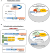
Model of MNDA nuclear function, cleavage and cytoplasmic relocalization. MNDA’s cellular localization and function vary in response to endogenous and exogenous stress signals. (A) Under normal conditions (resting state), MNDA’s PYD and HIN-200 (HIN-A) domains form an intramolecular association that results in a self-stabilized “resting” state, with the flexible link of the intrinsically disordered region (IDR) buckling. DNA binding to the HIN-200 domain and/or RNA association with the IDR or HIN-200 domain in the nucleoplasm weaken this intramolecular interaction and release constraints on the PYD signalling domain. The latter can then engage in homotypic or non-homotypic PYD-PYD interactions, for example with the nuclear protein ASC (apoptosis-associated speck-like protein containing a CARD) (not shown). MNDA can also associate with various nuclear proteins (86), including activating or repressive components of the positive transcription elongation factor b (P-TEFb) complex (43). In this “resting” state, MNDA could restrict the release of the promoter-proximal paused RNA polymerase II (Pol II) and, consequently, negatively affect transcriptional elongation of these genes. Grey oval indicated as ‘intx’ is a representation of the interaction between the PYD and HIN-200 (HIN-A) domains of MNDA. (B) In response to genotoxic or inflammation mediators such as danger- or pathogen-associated molecular patterns (DAMPs or PAMPs) (activated state), PYD-PYD interactions between MNDA and ASC proteins become possible, triggering inflammasome formation and, consequently, activation of pro-caspase-1. Among other substrates, catalytically active caspase-1 might induce proteolytic cleavage of MNDA. This would generate at least two characteristic MNDA fragments (43, 90). While the PYD-only fragment relocalizes to the cytoplasm where it may facilitate inflammasome assembly, the ‘IDR-HIN’ fragment would accumulate in the nucleus. Although it is not known whether the absence of PYD weakens the interaction of MNDA-HIN with repressive P-TEFb components such as HEXIM1, it has been shown that MNDA-dependent repression of transcription elongation is relieved following genotoxic stress (43). At the same time, MNDA may help stabilize newly transcribed RNA. This dual role of MNDA and the effect of MNDA on transcription elongation of MNDA targets, such as MCL1 and BCL2, could therefore be modulated by signals induced by genotoxic and inflammatory mediators.
Figure 4
Figure 4
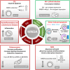
MNDA expression levels in human diseases. Schematic overview of the role of MNDA expression in human diseases and cellular functions. -In macrophages, MNDA can inhibit the DNA binding of the transcription factor Sp1 and thereby, blocks virus replication during human immunodeficiency virus 1 (HIV-1) infection (13). -Upon genotoxic stress in chronic lymphocytic leukaemia (CLL) cells or polymorphonuclear (PMN) leukocytes, MNDA represses MCL1 and BCL2 gene expression and enhances MCL-1 and BCL-2 protein degradation, promoting stress-induced apoptosis (90). -In granulocyte-monocyte progenitor (GMP) cells, reduced MNDA protein levels is associated with abnormal cell death and myeloid differentiation, and has been documented in familial (133) and sporadic (–135) myelodysplastic syndromes (MDS). MNDA binds nucleophosmin 1 (NPM1) and the NPM-MLF1 (myeloid leukaemia factor 1) chimera product (99), which has been associated with MDS (136). Whether these associations alter trafficking signals and/or direct inappropriate cellular localization of the aforementioned proteins in MDS cells remains to be elucidated. MNDA also interacts with nucleolin (NCL) and Yin Yang 1 (YY1) proteins, which are involved in cell cycle regulation and genotoxic stress response (137, 138), suggesting a possible role of MNDA in the control of MDS cell survival. In accordance with this data and regardless the fact that the precise mechanism of action is still undefined, MNDA expression was proposed as an independent clinical marker for the evaluation of dyspoiesis (139). -In osteosarcoma cells, the Hsa-miR-889-3p microRNA, whose upregulation is a known prognostic risk factor (130), inhibits MNDA expression whereas MNDA overexpression counteracts proliferation, induces apoptosis and reduces migration and invasiveness in the SAOS-2 osteosarcoma model cell line (131). -MNDA overexpression has been documented in monocytes obtained from individuals diagnosed with autoinflammatory (atherosclerotic lesions) (126); autoimmune (pancreatic islets of Langerhans in type 1 diabetes) (129); chronic inflammatory diseases such as ulcerative colitis (127) and chronic obstructive pulmonary disease (128).

References

    1. Oronsky B, Caroen S, Reid T. What exactly is inflammation (and what is it not)? Int J Mol Sci. (2022) 23. doi: 10.3390/ijms232314905 - DOI - PMC - PubMed
    1. Fernandes Q, Inchakalody VP, Bedhiafi T, Mestiri S, Taib N, Uddin S, et al. . Chronic inflammation and cancer; the two sides of a coin. Life Sci. (2024) 338:122390. doi: 10.1016/j.lfs.2023.122390 - DOI - PubMed
    1. Alfaro S, Acuna V, Ceriani R, Cavieres MF, Weinstein-Oppenheimer CR, Campos-Estrada C. Involvement of inflammation and its resolution in disease and therapeutics. Int J Mol Sci. (2022) 23. doi: 10.3390/ijms231810719 - DOI - PMC - PubMed
    1. Milot E, Fotouhi-Ardakani N, Filep JG. Myeloid nuclear differentiation antigen, neutrophil apoptosis and sepsis. Front Immunol. (2012) 3:397. doi: 10.3389/fimmu.2012.00397 - DOI - PMC - PubMed
    1. Kakiuchi T, Nariuchi H, Matsuhashi T. Fc receptors on IgM and IgG antibody-forming cells in an early immune response. Cell Immunol. (1981) 61:124–33. doi: 10.1016/0008-8749(81)90359-2 - DOI - PubMed

Publication types

Substances

LinkOut - more resources