. 2024 Apr 12:15:1373745.
doi: 10.3389/fimmu.2024.1373745. eCollection 2024.

Myeloid A20 is critical for alternative macrophage polarization and type-2 immune-mediated helminth resistance

Affiliations

Myeloid A20 is critical for alternative macrophage polarization and type-2 immune-mediated helminth resistance

Ioanna Petta et al. Front Immunol. .

Abstract

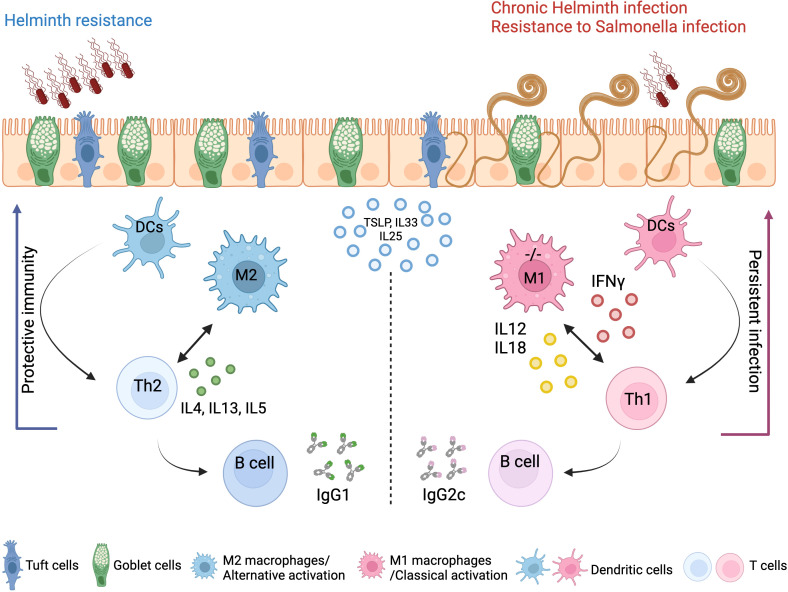
Background: Protective immunity against intestinal helminths requires induction of robust type-2 immunity orchestrated by various cellular and soluble effectors which promote goblet cell hyperplasia, mucus production, epithelial proliferation, and smooth muscle contractions to expel worms and re-establish immune homeostasis. Conversely, defects in type-2 immunity result in ineffective helminth clearance, persistent infection, and inflammation. Macrophages are highly plastic cells that acquire an alternatively activated state during helminth infection, but they were previously shown to be dispensable for resistance to Trichuris muris infection.

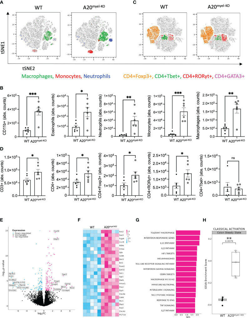
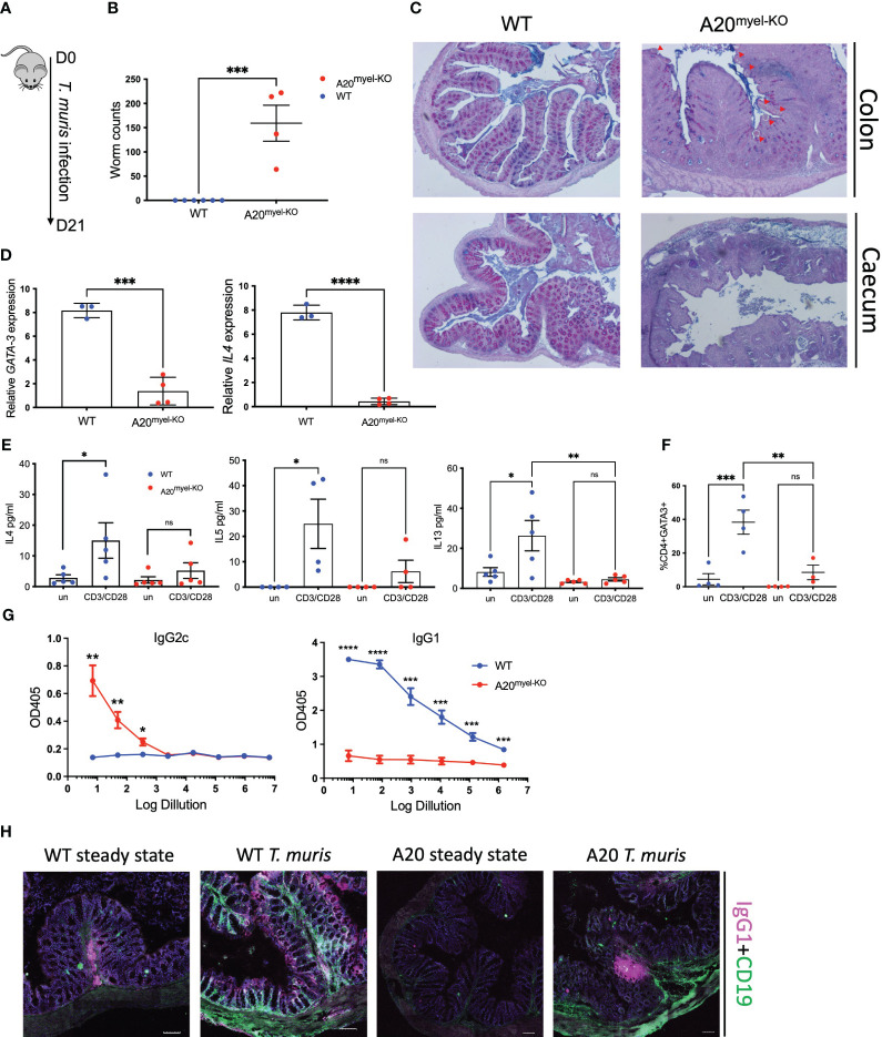
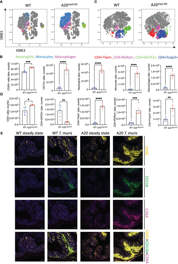
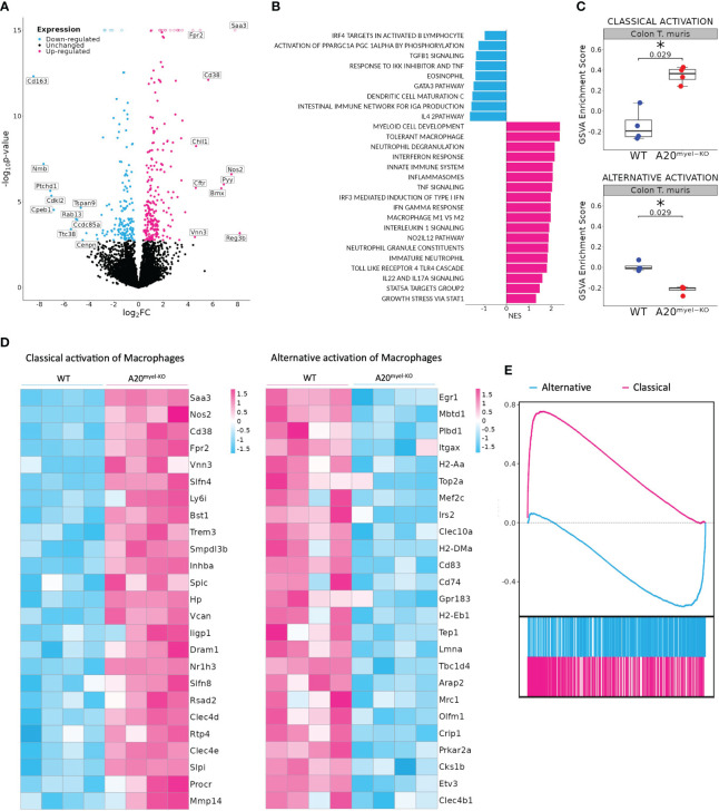
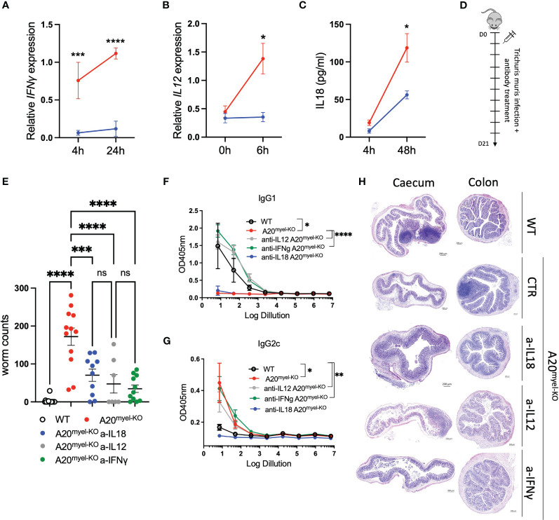
Methods: We use the in vivo mouse model A20myel-KO, characterized by the deletion of the potent anti-inflammatory factor A20 (TNFAIP3) specifically in the myeloid cells, the excessive type-1 cytokine production, and the development of spontaneous arthritis. We infect A20myel-KO mice with the gastrointestinal helminth Trichuris muris and we analyzed the innate and adaptive responses. We performed RNA sequencing on sorted myeloid cells to investigate the role of A20 on macrophage polarization and type-2 immunity. Moreover, we assess in A20myel-KO mice the pharmacological inhibition of type-1 cytokine pathways on helminth clearance and the infection with Salmonella typhimurium.

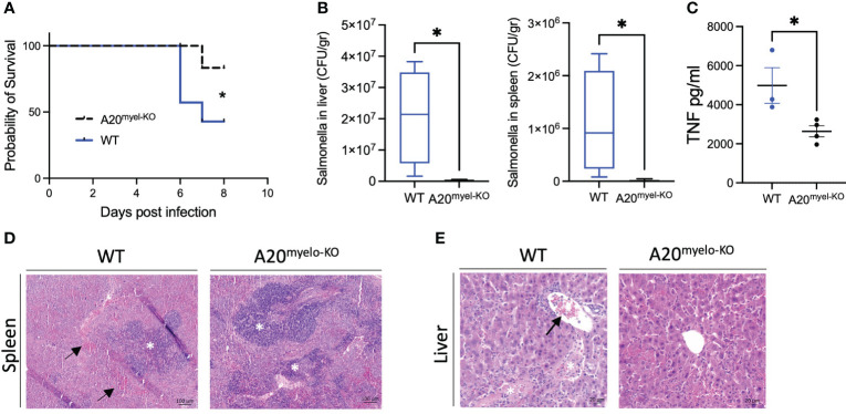
Results: We show that proper macrophage polarization is essential for helminth clearance, and we identify A20 as an essential myeloid factor for the induction of type-2 immune responses against Trichuris muris. A20myel-KO mice are characterized by persistent Trichuris muris infection and intestinal inflammation. Myeloid A20 deficiency induces strong classical macrophage polarization which impedes anti-helminth type-2 immune activation; however, it promotes detrimental Th1/Th17 responses. Antibody-mediated neutralization of the type-1 cytokines IFN-γ, IL-18, and IL-12 prevents myeloid-orchestrated Th1 polarization and re-establishes type-2-mediated protective immunity against T. muris in A20myel-KO mice. In contrast, the strong Th1-biased immunity in A20myel-KO mice offers protection against Salmonella typhimurium infection.

Conclusions: We hereby identify A20 as a critical myeloid factor for correct macrophage polarization and appropriate adaptive mucosal immunity in response to helminth and enteric bacterial infection.

Keywords: A20 (TNFAIP3); adaptive immunity; helminth infection; immunity to parasites; innate immunity; intestinal immunity; macrophage polarization; type-2 response.


Conflict of interest statement

Author LB is employed by the company JJP Biologics. The remaining authors declare that the research was conducted in the absence of any commercial or financial relationships that could be construed as a potential conflict of interest.

Figures

Figure 1
Figure 1
A20 deletion in myeloid cells affects immune cell composition and favors M1 macrophage polarization. (A, B) Flow cytometry analysis of colon lamina propria of WT (n = 6) and A20myel-KO (n = 6) mice for the myeloid and (C, D) T-cell composition, presented in t-SNE plots (upper panels) and bar graphs (lower panels). All data are presented as means ± s.e.m. Statistical significance was determined by a two-sided Student’s t-test. (E) Bulk RNA sequencing volcano plots presenting the differentially expressed genes in CD11b+ sorted cells of WT (n = 5, blue) and A20myel-KO (n = 5, magenta) colon samples. (F) Heatmap showing the top 25 M1 markers to be upregulated in A20myel-KO colon compared with WT. (G) Gene ontology pathway analysis showing the positively regulated pathways in A20meyl-KO colon CD11b+ cells. (H) Gene scoring analysis using an M1 signature consisting of 700 genes to compare the expression in WT and A20myel-KO colon. * p<0,05, ** p<0,01, *** p<0,001. Ns, Not significant.
Figure 2
Figure 2
A20myel-KO mice are susceptible to Trichuris muris infection. (A) Infection scheme with Trichuris muris gastrointestinal helminth. (B) Worm counts in the cecum of WT (n = 6, blue) and A20myel-KO (n = 4, red) 21 days after infection with T. muris. (C) Representative images of AB/PAS staining of WT and A20myel-KO colon (upper panel) and cecum (lower panel) 21 days after T. muris infection. Scale bars 100 μm. (D) GATA3 and IL-4 relative expression in WT (n = 3) and A20myel-KO (n = 4) total colon lysates. (E) Cytokine levels of IL-4, IL-5, and IL-13 in the supernatant restimulated with a-CD3/a-CD28 WT and A20myel-KO mLN for 48 h. (F) CD4+GATA3+ levels determined by flow cytometry analysis in restimulated mLN and of WT and A20myel-KO. (G) ELISA for the measurement of IgG2c and IgG1 levels in the blood serum of WT and A20myel-KO mice infected with T. muris for 21 days. (H) MACSima imaging for IgG1 (magenta) and CD19 (green) of WT and A20myel-KO colon sections at steady state and upon infection with T. muris for 21 days. Scale bars 100 μm. All data are presented as means ± s.e.m. Statistical significance was determined by a two-sided Student’s t-test or by one-way ANOVA for multiple comparisons. * p<0,05, ** p<0,01, *** p<0,001, **** p < 0.0001. Ns, Not significant.
Figure 3
Figure 3
Deletion of A20 in myeloid cells hampers Th2 immunity and promotes Th1 response upon Trichuris muris infection. (A, B) Flow cytometry analysis of colon lamina propria of WT (n = 6) and A20myel-KO (n = 4) mice infected with T. muris for the myeloid and (C, D) T-cell composition, presented in t-SNE plots (upper panels) and bar graphs (lower panels). (E) MACSima fluorescent imaging for CD45 (yellow), CD11b (green), and CD64 (magenta) of WT and A20myel-KO colon sections at steady state and upon infection with T. muris for 21 days. Scale bar 100 μm. * p<0,05, ** p<0,01, *** p<0,001, **** p < 0.0001.
Figure 4
Figure 4
A20myel-KO are characterized by M1 macrophage signature upon Trichuris muris infection. (A) Bulk RNA sequencing volcano plots presenting the differentially expressed genes in CD11b+ sorted cells of WT (blue) and A20myel-KO (magenta) colon samples of mice infected with T. muris for 10 days. (B) Gene ontology pathway analysis showing the positively regulated pathways in WT (blue) and A20meyl-KO (magenta) colon CD11b+ cells of mice infected with T. muris for 10 days. (C) Gene scoring analysis using M1 and M2 signatures, consisting of 710 and 806 genes, respectively, to compare the expression in WT and A20myel-KO colon upon T. muris infection. (D) Heatmap showing the upregulation of the top 25 markers related to classical activation of macrophages (M1) in A20myel-KO mice compared with WT (left). Heatmap showing the upregulation of the top 25 markers related to the alternative activation of macrophages (M2) in WT compared with A20myel-KO mice (right). (E) Gene set enrichment analysis (GSEA) plots visualizing the enrichment of classical (M1, upper panel) and alternative (M2, lower panel) activation of macrophages in A20myel-KO and WT, respectively. Each plot features the running enrichment score (y-axis) and shows the placement of the ranked differentially expressed genes for the respective transcriptomic signature (x-axis).
Figure 5
Figure 5
Blocking Th1-related cytokine reverse susceptibility of A20myel-KO to Trichuris muris. (A) Relative IFN-γ expression in BMDMs derived from WT (blue) and A20myel-KO (red) stimulated with Trichuris antigen for 4 h and 24 h. (B) Relative expression of IL-12 in WT (blue) and A20myel-KO (red)-derived BMDMs stimulated for 0 h and 6 h with T. muris antigen. (C) IL-18 levels in the supernatant of WT (blue) and A20myel-KO (red)-derived BMDMs, stimulated with Trichuris antigen for 4 h and 48 h. (D) Infection and treatment scheme with T. muris and neutralizing antibodies against IFN-γ, IL-12, and IL-18. (E) Worm counts in the cecum of WT (n = 15, open dots), A20myel-KO+IgG control antibody (n = 12, red), A20myel-KO+a-IL-18 (n = 10, blue), A20myel-KO+a-IL-12 (n = 7, gray), and A20myel-KO+a-IFN-γ (n = 11, green) 21 days after infection with T. muris. The graph represents the combined data of three independent experiments. (F) ELISA for IgG1 and (G) IgG2c levels in the blood serum of WT and A20myel-KO mice treated as in (B). Significance refers to log dilution 1. (H) Representative images of AB/PAS staining of cecum and colon sections of WT and A20myel-KO mice infected with T. muris and treated with neutralizing antibodies, as in (B). Scale bars 200 μm. Data are presented as means ± s.e.m. Statistical significance was determined by a two-sided Student’s t-test or by one-way ANOVA for multiple comparisons. * p<0,05, ** p<0,01, *** p<0,001, **** p < 0.0001. Ns, Not significant.
Figure 6
Figure 6
A20myel-KO mice are protected against Salmonella typhimurium infection. (A) Survival curve of WT (n = 7) and A20myel-KO (n = 6) mice during 8 days of infection with S. typhimurium. Statistical analysis is performed using the Gehan–Breslow–Wilcoxon test. (B) CFU counts of retrieved S. typhimurium from the liver and spleen of infected WT (n = 4) and A20myel-KO (n = 5) 8 days post-infection. (C) ELISA for TNF levels in the blood serum of WT (n = 4) and A20myel-KO (n = 5) 8 days post-infection. Data are presented as means ± s.e.m. Statistical significance was determined by a two-sided Student’s t-test. (D) Representative images of H&E staining of spleens of infected WT and A20myel-KO mice. The black arrows indicate hematopoiesis (red pulp) and the white asterisks indicate the lymphoid tissue (white pulp). Scale bar 100 μm. (E) Representative images of H&E staining of the liver of infected WT and A20myel-KO mice. The black arrow indicates immune cell infiltration and the white asterisks indicate the necrotic areas. Scale bar 20 μm. * p<0,05.
Figure 7
Figure 7
Concluding scheme. The clearance of gastrointestinal helminths depends on type-2 immunity. Helminths interact with and damage intestinal tissue, which leads to the release of intracellular DAMPs and cytokines such as TSLP and IL-33 and IL-25 produced by epithelial cells. These factors activate myeloid cells, which further activate T and B cells to mount effective Th2 responses and the secretion of IL-4, IL-5, and IL-13 cytokines, as well as helminth-specific IgG1 immunoglobulins, leading to effective expulsion of the helminths. Deletion of A20 in the myeloid cells leads to a strong classical macrophage polarization and enhanced secretion of type-1-associated cytokines, including IL-12, IL-18, and IFN-γ, which impede type-2 immune-mediated helminth clearance and promotes persistent infection and intestinal inflammation. Created with BioRender.com.

Similar articles

Cited by

References

    1. WHO . Investing to overcome the global impact of neglected tropical diseases: third WHO report on neglected diseases. (2015) Geneva, Switzerland: WHO Press, World Health Organization.
    1. Loukas A, Maizels RM, Hotez PJ. The yin and yang of human soil-transmitted helminth infections. Int J Parasitol. (2021) 51:1243–53. doi: 10.1016/j.ijpara.2021.11.001 - DOI - PMC - PubMed
    1. Pullan RL, Smith JL, Jasrasaria R, Brooker SJ. Global numbers of infection and disease burden of soil transmitted helminth infections in 2010. Parasit Vectors. (2014) 7. doi: 10.1186/1756-3305-7-37 - DOI - PMC - PubMed
    1. Colombo SAP, Grencis RK. Immunity to soil-transmitted helminths: evidence from the field and laboratory models. Front Immunol. (2020) 11:1286. doi: 10.3389/fimmu.2020.01286 - DOI - PMC - PubMed
    1. Lechner A, Bohnacker S, Esser-von Bieren J. Macrophage regulation & function in helminth infection. Semin Immunol. (2021) 53. doi: 10.1016/j.smim.2021.101526 - DOI - PubMed

Publication types

Substances