. 2023 May 10;9(4):359-368.
doi: 10.1002/ibra.12103. eCollection 2023 Winter.

Ultrasound-guided puncture into newborn rat brain

Affiliations

Ultrasound-guided puncture into newborn rat brain

Rui-Fang Ma et al. Ibrain. .

Abstract

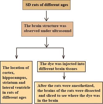
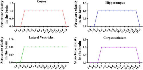
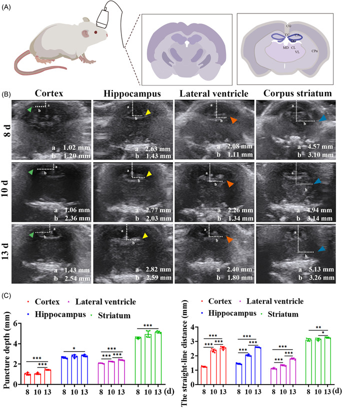
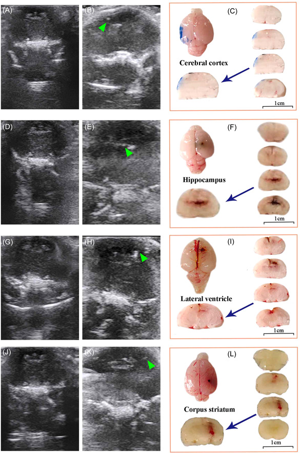
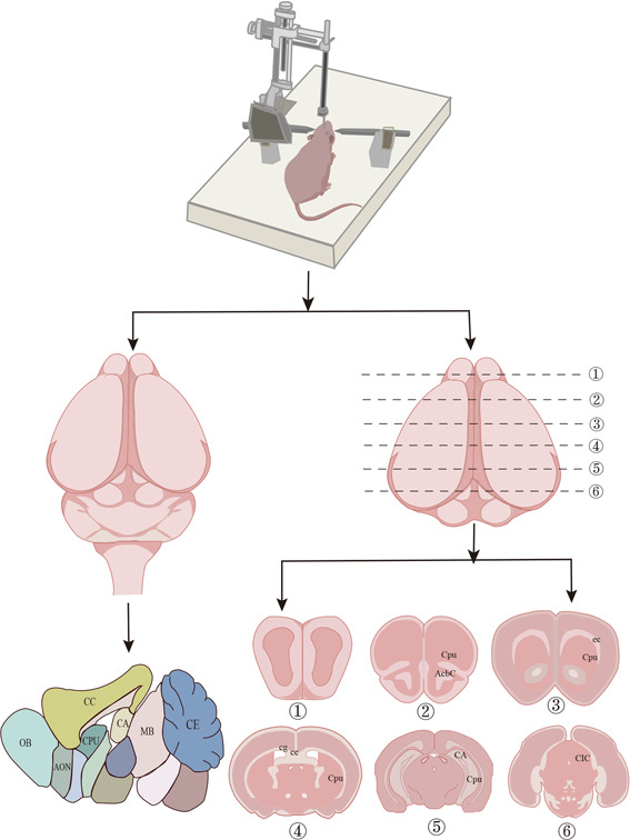
Since the brain structure of neonatal rats was not fully formed during the first 4 days, it cannot be detected using ultrasound. The objective of this study was to investigate the use of ultrasound to guide puncture in the normal coronal brain structure and determine the puncture depth of the location of the cortex, hippocampus, lateral ventricle, and striatum of newborn rats of 5-15 days. The animal was placed in a prone position. The specific positions of the cortex, hippocampus, lateral ventricle, and striatum were measured under ultrasound. Then, the rats were punctured with a stereotaxic instrument, and dye was injected. Finally, the brains of rats were taken to make frozen sections to observe the puncture results. By ultrasound, the image of the cortex, hippocampus, lateral ventricle, and striatum of the rat can be obtained and the puncture depth of the cortex (8 days: 1.02 ± 0.12, 10 days: 1.02 ± 0.08, 13 days: 1.43 ± 0.05), hippocampus (8 days: 2.63 ± 0.07, 10 days: 2.77 ± 0.14, 13 days: 2.82 ± 0.09), lateral ventricle (8 days: 2.08 ± 0.04, 10 days: 2.26 ± 0.03, 13 days: 2.40 ± 0.06), and corpus striatum (8 days: 4.57 ± 0.09, 10 days: 4.94 ± 0.31, 13 days: 5.13 ± 0.10) can be accurately measured. The rat brain structure and puncture depth changed with the age of the rats. Ultrasound technology can not only clarify the brain structure characteristics of 5-15-day-old rats but also guide the puncture and injection of the rat brain structure. The results of this study laid the foundation for the future use of ultrasound in experimental animal models of neurological diseases.

Keywords: brain structure; cortex; hippocampus; lateral ventricle; striatum; ultrasonoscopy.


Conflict of interest statement

Kun Zhang is employed by the company Shantou Ultrasonic Instrument Research Institute Co. Ltd. He confirms that he has no commercial interest that could be produced from this manuscript. The remaining authors declare that the research was conducted in the absence of any commercial or financial relationships that could be construed as a potential conflict of interest.

Figures

Figure 1
Figure 1
Study flowchart. To determine the location of the cortex, hippocampus, lateral ventricle, and striatum of rats of different ages, the location of the brain structure of rats was detected by ultrasound. The puncture was performed under the ultrasonic locator. After the dye was injected into the cortex, hippocampus, lateral ventricle, and striatum, the brain tissue was taken after anesthetizing the rats, frozen sections were made, and the puncture location was compared. [Color figure can be viewed at wileyonlinelibrary.com]
Figure 2
Figure 2
Coronal dynamic exploration of rat brain structure clarity. Coronal ultrasound was used to detect the structural clarity of the cerebral cortex, hippocampus, lateral ventricle, and corpus striatum; 1 represents clear brain structure and 0 means that the brain structure is not clear. d, days. [Color figure can be viewed at wileyonlinelibrary.com]
Figure 3
Figure 3
Ultrasound‐guided localization of puncture in different brain regions. (A) Schematic diagram of the brain structure of rats under ultrasound. (B) Cortex, hippocampus, lateral ventricle, and striatum images of normal rats under ultrasound. a Puncture depth. b Straight‐line distance from the anterior midline to the puncture point. 8, 10, and 13 d represent the age of normal rats. The green arrow points to the cortex; the yellow arrow points to the hippocampus; the red arrow points to the lateral ventricle; and the blue arrow points to the striatum. (C) Quantified data including the puncture depth and the straight‐line distance from the anterior midline to the puncture point. CA: hippocampus; CC: cerebral cortex; CG: cingulum; CL: centrolateral thalamic nucleus; CM: central medial thalamic nucleus; Cpu: caudate putamen; d: days; DG: dentate gyrus; MD: mediodorsal thalamic nucleus; VL: ventrolateral thalamic nucleus; VPM: ventral posterolateral thalamic nucleus. *p < 0.05; **p < 0.01; ***p < 0.001. [Color figure can be viewed at wileyonlinelibrary.com]
Figure 4
Figure 4
Localization images of the cortex, hippocampus, lateral ventricle, and striatum in the rat brain structure under ultrasound. (A, D, G, L) Coronal images of rats under ultrasound; images of the cortex (B), hippocampus (E), lateral ventricles (H), and striatum (M); coronal images of the brain section, dye‐stained position: cortex (C), hippocampus (F), lateral ventricle (I), and striatum (N). The scale bar of the brain section is 1 cm. [Color figure can be viewed at wileyonlinelibrary.com]
Figure 5
Figure 5
Example diagram of the research. Anatomy under ultrasound‐guided puncture. The research route of the cortex, hippocampus, lateral ventricle, and striatum puncture positions of rats of different ages under ultrasound. ①–⑥ represent coronal sections of brain anatomy in SD rats. AcbC: accumbens nucleus, core; AON: ameror olfactory nucleus; CA: hippocampus; CC: cerebral cortex; CE: central amygdaloid nucleus; cg: cingulum; CIC: Central nucleus of inferior colliculus; Cpu: caudate putamen; MB: mamillary body; OB: Olfactory Bulb. [Color figure can be viewed at wileyonlinelibrary.com]

Similar articles

References

    1. Hakimi R, Alexandrov AV, Garami Z. Neuro‐ultrasonography. Neurol Clin. 2020;38(1):215‐229. 10.1016/j.ncl.2019.09.006 - DOI - PubMed
    1. Dietrich C, van Lieshout J, Fischer I, et al. Transcranial Doppler ultrasound, perfusion computerized tomography, and cerebral angiography identify different pathological entities and supplement each other in the diagnosis of delayed cerebral ischemia. Acta Neurochir Suppl. 2020;127:155‐160. 10.1007/978-3-030-04615-6_23 - DOI - PubMed
    1. Schultz KL, Quinn SM, Miller AH, et al. Point of care ultrasound facilitated diagnosis of right ventricular mass as the etiology of syncope; A case report of intravenous leiomyomatosis. Radiol Case Rep. 2021;16(6):1288‐1293. 10.1016/j.radcr.2021.02.062 - DOI - PMC - PubMed
    1. Weile J, Frederiksen CA, Laursen CB, Graumann O, Sloth E, Kirkegaard H. Point‐of‐care ultrasound induced changes in management of unselected patients in the emergency department ‐ a prospective single‐blinded observational trial. Scand J Trauma Resusc Emerg Med. 2020;28(1):47. 10.1186/s13049-020-00740-x - DOI - PMC - PubMed
    1. Lau VI, Jaidka A, Wiskar K, et al. Better with ultrasound. Chest. 2020;157(1):142‐150. 10.1016/j.chest.2019.08.2204 - DOI - PubMed