Comparative Study
. 2024 Apr 14;30(14):2038-2058.
doi: 10.3748/wjg.v30.i14.2038.

Comparative transcriptomic analysis reveals the molecular changes of acute pancreatitis in experimental models

Affiliations
Comparative Study

Comparative transcriptomic analysis reveals the molecular changes of acute pancreatitis in experimental models

Pan Zheng et al. World J Gastroenterol. .

Abstract

Background: Acute pancreatitis (AP) encompasses a spectrum of pancreatic inflammatory conditions, ranging from mild inflammation to severe pancreatic necrosis and multisystem organ failure. Given the challenges associated with obtaining human pancreatic samples, research on AP predominantly relies on animal models. In this study, we aimed to elucidate the fundamental molecular mechanisms underlying AP using various AP models.

Aim: To investigate the shared molecular changes underlying the development of AP across varying severity levels.

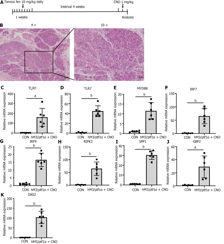
Methods: AP was induced in animal models through treatment with caerulein alone or in combination with lipopolysaccharide (LPS). Additionally, using Ptf1α to drive the specific expression of the hM3 promoter in pancreatic acinar cells transgenic C57BL/6J- hM3/Ptf1α(cre) mice were administered Clozapine N-oxide to induce AP. Subsequently, we conducted RNA sequencing of pancreatic tissues and validated the expression of significantly different genes using the Gene Expression Omnibus (GEO) database.

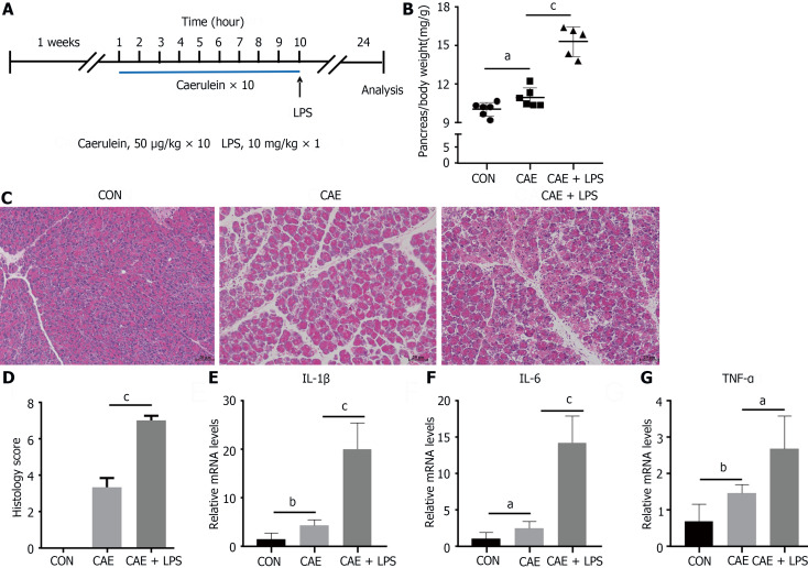
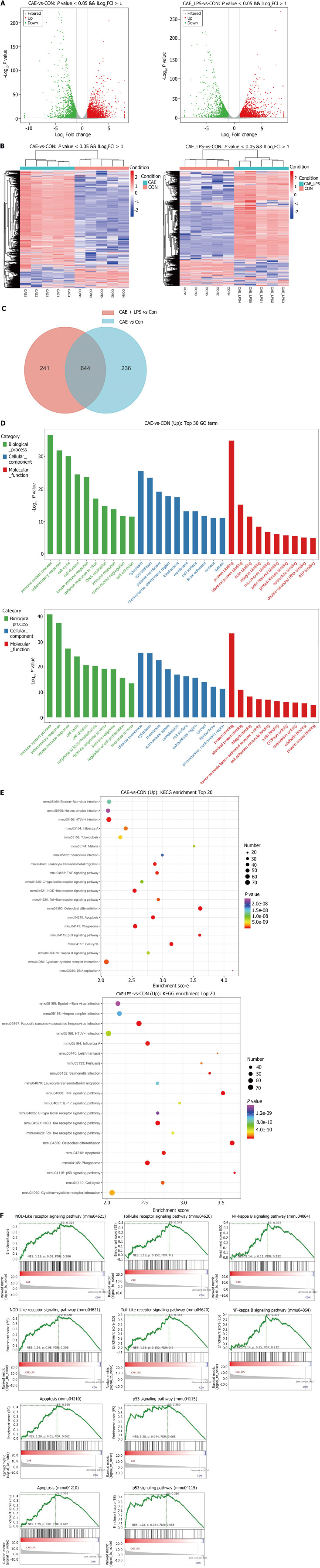
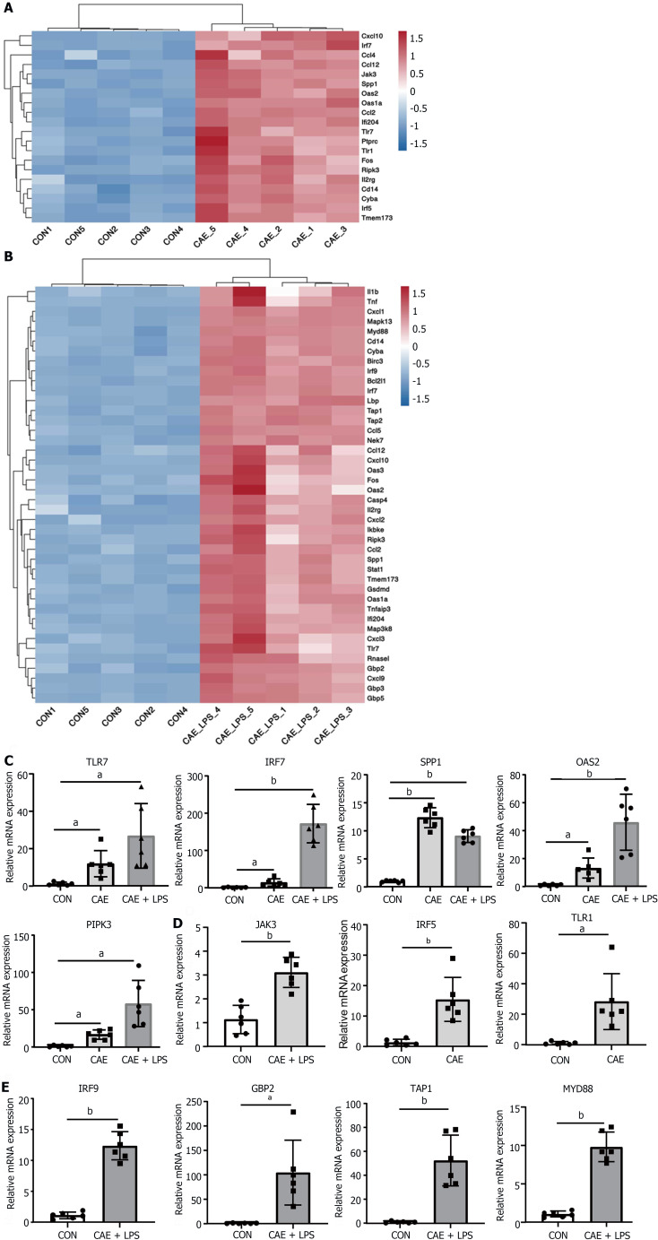
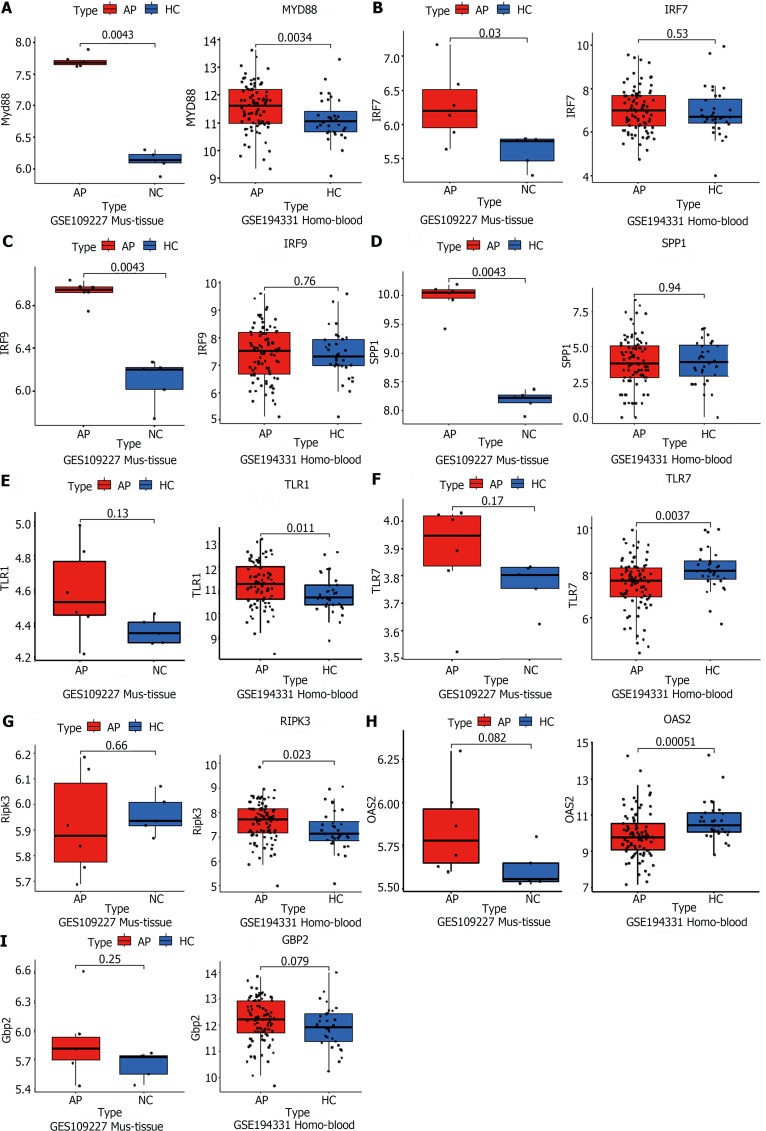
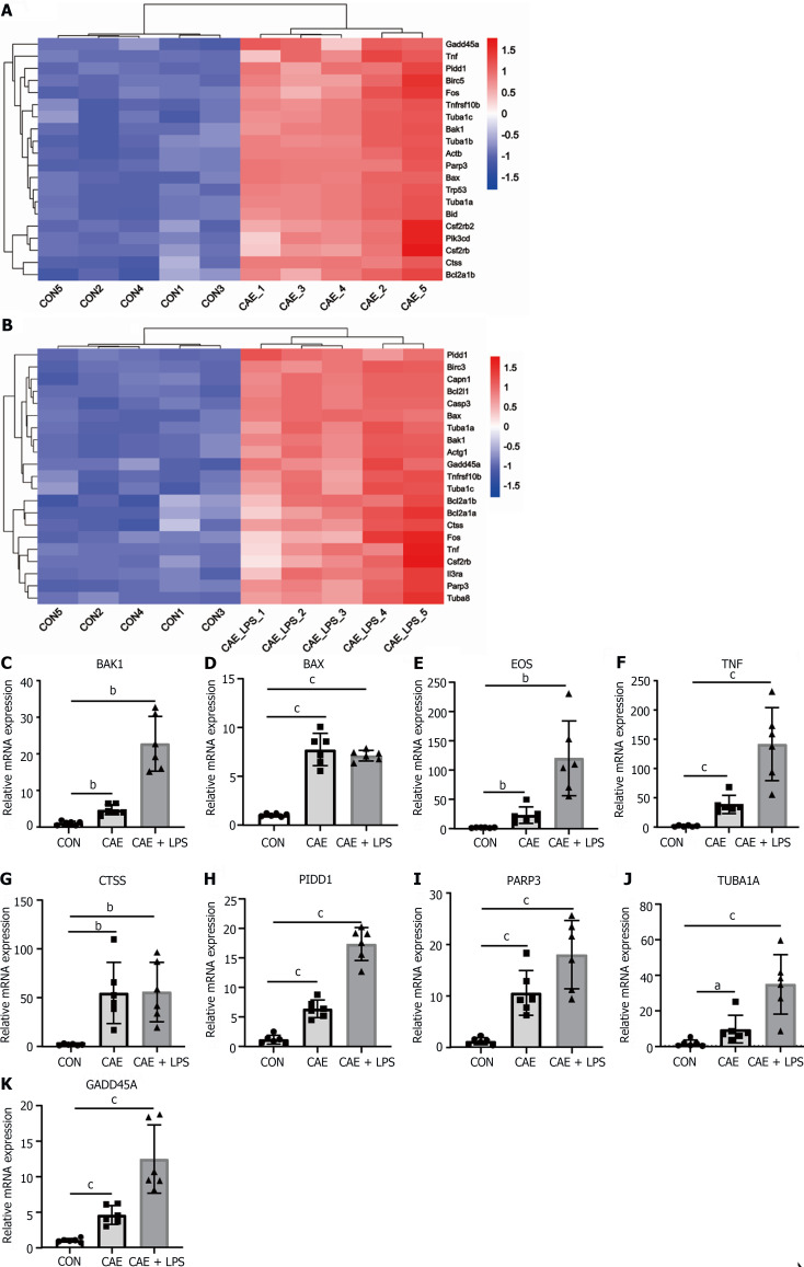
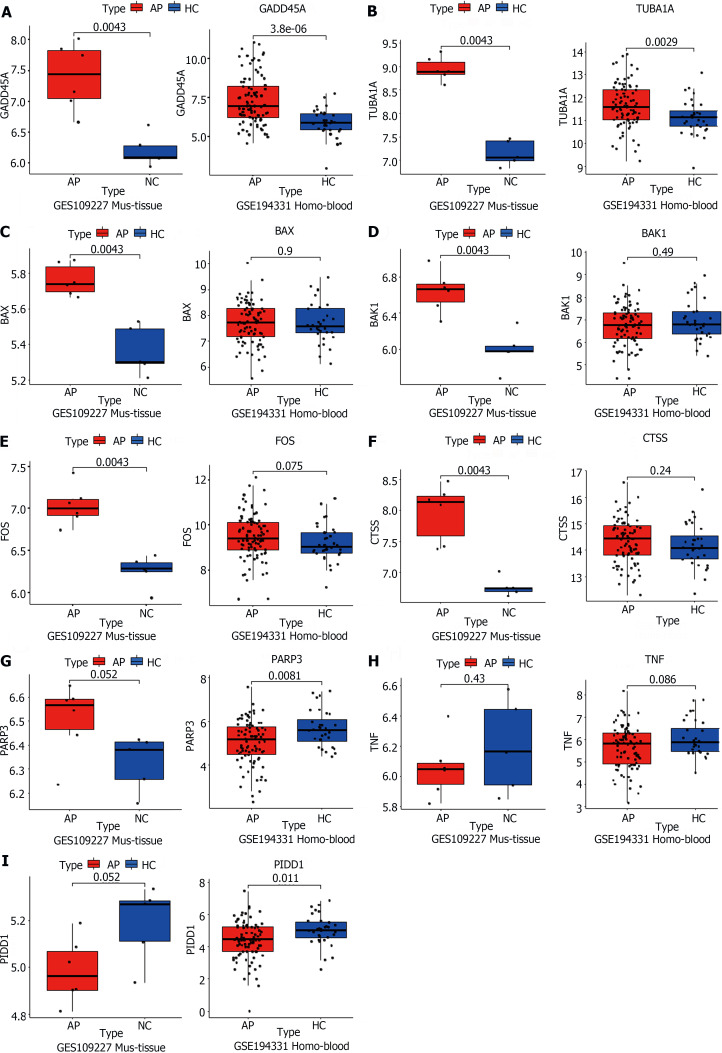
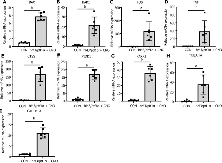
Results: Caerulein-induced AP showed severe inflammation and edema, which were exacerbated when combined with LPS and accompanied by partial pancreatic tissue necrosis. Compared with the control group, RNA sequencing analysis revealed 880 significantly differentially expressed genes in the caerulein model and 885 in the caerulein combined with the LPS model. Kyoto Encyclopedia of Genes and Genomes enrichment analysis and Gene Set Enrichment Analysis indicated substantial enrichment of the TLR and NOD-like receptor signaling pathway, TLR signaling pathway, and NF-κB signaling pathway, alongside elevated levels of apoptosis-related pathways, such as apoptosis, P53 pathway, and phagosome pathway. The significantly elevated genes in the TLR and NOD-like receptor signaling pathways, as well as in the apoptosis pathway, were validated through quantitative real-time PCR experiments in animal models. Validation from the GEO database revealed that only MYD88 concurred in both mouse pancreatic tissue and human AP peripheral blood, while TLR1, TLR7, RIPK3, and OAS2 genes exhibited marked elevation in human AP. The genes TUBA1A and GADD45A played significant roles in apoptosis within human AP. The transgenic mouse model hM3/Ptf1α(cre) successfully validated significant differential genes in the TLR and NOD-like receptor signaling pathways as well as the apoptosis pathway, indicating that these pathways represent shared pathological processes in AP across different models.

Conclusion: The TLR and NOD receptor signaling pathways play crucial roles in the inflammatory progression of AP, notably the MYD88 gene. Apoptosis holds a central position in the necrotic processes of AP, with TUBA1A and GADD45A genes exhibiting prominence in human AP.

Keywords: Acute pancreatitis; Apoptosis; Experimental acute pancreatitis models; Inflammatory; RNA-sequencing; TLR and NOD-like signaling pathways.


Conflict of interest statement

Conflict-of-interest statement: The authors declare that they have no competing interests.

Figures

Figure 1
Figure 1
Treatment with caerulein alone or in combination with lipopolysaccharide induced varying degrees of experimental acute pancreatitis. Modeling methods for the control group, caerulein (CAE) group, and CAE combined with lipopolysaccharide (LPS; CAE + LPS) group, evaluation of pancreatic tissue changes, and relevant parameters. A: Schematic representation of the modeling methods for the CAE group and CAE + LPS group; B: Pancreatic weight ratios in the three groups; C: H&E staining of pancreatic tissue; D: Scores for edema, inflammation, and necrosis in pancreatic pathology; E-G: Quantitative real-time PCR expression values of pancreatic inflammatory factors. n = 6 per group, aP < 0.05, bP < 0.01, cP < 0.001. CAE: Caerulein; CON: Control; LPS: Lipopolysaccharide.
Figure 2
Figure 2
RNA-seq identified the molecular changes during the development of acute pancreatitis. Comparative analysis of the caerulein (CAE) group and CAE + lipopolysaccharide (LPS) group with the control (Con) group. A: Volcano plot of significant differentially expressed (SDE) genes (P value < 0.05 and |log2FC| > 1), with red indicating significant upregulation and green indicating significant downregulation; B: Venn diagram, where blue represents CAE group vs Con group, and red represents CAE + LPS group vs Con group, with the gray area representing commonly differentially expressed genes in both groups compared to the Con group; C: Heatmap displaying the expression of SDE genes in each pancreatic sample, with red indicating upregulated genes and blue indicating downregulated genes; D: Gene Ontology (GO) functional annotation of the top 30 significantly different genes, with the GO classification chart showing the distribution of entries related to biological processes, cellular components, and molecular functions; E: Enrichment analysis results of the top 20 significantly different Kyoto Encyclopedia of Genes and Genomes pathways; F: Gene Set Enrichment Analysis for the NOD-like receptor, TLR, NF-κB, and P53 signaling pathways, as well as apoptosis. CAE: Caerulein; CON: Control; LPS: Lipopolysaccharide.
Figure 3
Figure 3
NOD-like receptor and TLR signaling pathway played a crucial role in the progression of acute pancreatitis. Significant differentially expressed genes in the NOD-like receptor and TLR signaling pathways after comparing the caerulein (CAE) group and CAE + lipopolysaccharide (LPS) group to the control (Con) group. A: Heatmap of significant differentially expressed (SDE) genes in the CAE Group compared to the Con group; B: Heatmap of SDE genes in the CAE + LPS group compared to the Con group; C: Quantitative real-time PCR (qRT-PCR) expression values of commonly significantly different genes in both groups; D: qRT-PCR expression values of specifically significantly different genes in the CAE Group compared to the Con group; E: qRT-PCR expression values of specifically significantly different genes in the CAE + LPS group compared to the Con group. n = 6 per group, aP < 0.01, bP < 0.001. CAE: Caerulein; CON: Control; LPS: Lipopolysaccharide.
Figure 4
Figure 4
Public RNA-seq data reveals that the significant role of TLR and NOD-like signaling pathways in acute pancreatitis pathogenesis. A-I: The acute pancreatitis (AP) animal RNA-seq dataset (GSE109227) and human AP patient blood RNA-seq dataset (GSE194331) were downloaded from the Gene Expression Omnibus database for the external validation of significant differentially expressed genes in the successfully validated NOD-like receptor and TLR signaling pathways in the animal model, using quantitative real-time PCR. n = 6 per group.
Figure 5
Figure 5
Apoptosis served as a crucial pathway mediating pancreatic necrosis in acute pancreatitis. Significant differentially expressed (SDE) genes in the apoptotic signaling pathway after comparing the caerulein (CAE) group and CAE + lipopolysaccharide (LPS) group to the control (Con) group. A: Heatmap of SDE genes in the CAE group compared to the Con group; B: Heatmap of SDE genes in the CAE + LPS group compared to the Con group; C-K: Quantitative real-time PCR expression values of commonly significantly different genes in both groups, n = 6 per group, aP < 0.05, bP < 0.01, cP < 0.001. CAE: Caerulein; CON: Control; LPS: Lipopolysaccharide.
Figure 6
Figure 6
TUBA1A and GADD45A were genes closely associated with apoptosis in acute pancreatitis. A-I: The acute pancreatitis (AP) animal RNA-seq dataset (GSE109227) and human AP patient blood RNA-seq dataset (GSE194331) were downloaded from the Gene Expression Omnibus database for the external validation of the genes successfully validated in the animal model and related to the apoptotic signaling pathway, using quantitative real-time PCR. AP: Acute pancreatitis; HC: Healthy control.
Figure 7
Figure 7
Inflammatory response in hM3/Ptf1α(cre) transgenic animal models consistent with the caerulein model. hM3/Ptf1α(cre) model and its pathological images with inflammatory gene validation. A: Schematic representation of the hM3/Ptf1α(cre) model; B: H&E staining of pancreatic tissue schematic representation of the modeling methods; C-K: Validation of the significant differentially expressed genes in the transgenic animal hM3/Ptf1α(cre) model after inducing acute pancreatitis, which were found in the NOD-like receptor and TLR signaling pathways. n = 6 per group, aP < 0.01, bP < 0.001. CON: Control; CNO: Clozapine N-oxide.
Figure 8
Figure 8
The apoptosis in acute pancreatitis in the hM3/Ptf1α(cre) transgenic animal model was consistent with the caerulein models. A-I: Validation of the significant differentially expressed genes in the NOD-like receptor and TLR signaling pathways in the transgenic animal hM3/Ptf1α(cre) model after inducing acute pancreatitis. n = 6 per group, aP < 0.01, bP < 0.001. CON: Control; CNO: Clozapine N-oxide.

Similar articles

Cited by

References

    1. Habtezion A, Gukovskaya AS, Pandol SJ. Acute Pancreatitis: A Multifaceted Set of Organelle and Cellular Interactions. Gastroenterology. 2019;156:1941–1950. - PMC - PubMed
    1. Garg PK, Singh VP. Organ Failure Due to Systemic Injury in Acute Pancreatitis. Gastroenterology. 2019;156:2008–2023. - PMC - PubMed
    1. Boxhoorn L, Voermans RP, Bouwense SA, Bruno MJ, Verdonk RC, Boermeester MA, van Santvoort HC, Besselink MG. Acute pancreatitis. Lancet. 2020;396:726–734. - PubMed
    1. Lampel M, Kern HF. Acute interstitial pancreatitis in the rat induced by excessive doses of a pancreatic secretagogue. Virchows Arch A Pathol Anat Histol. 1977;373:97–117. - PubMed
    1. Lerch MM, Gorelick FS. Models of acute and chronic pancreatitis. Gastroenterology. 2013;144:1180–1193. - PubMed

Publication types

MeSH terms