. 2024 Apr 23:18:1349-1368.
doi: 10.2147/DDDT.S458013. eCollection 2024.

Propofol and Dexmedetomidine Ameliorate Endotoxemia-Associated Encephalopathy via Inhibiting Ferroptosis

Affiliations

Propofol and Dexmedetomidine Ameliorate Endotoxemia-Associated Encephalopathy via Inhibiting Ferroptosis

Ye Zhou et al. Drug Des Devel Ther. .

Abstract

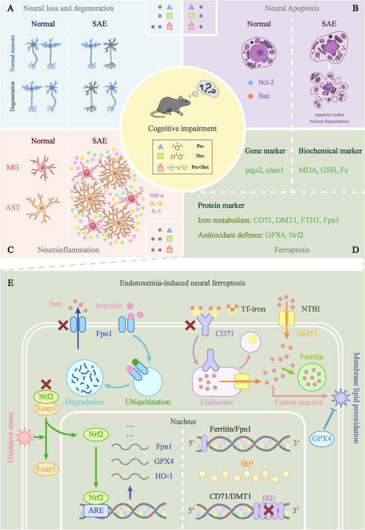
Background: Sepsis is recognized as a multiorgan and systemic damage caused by dysregulated host response to infection. Its acute systemic inflammatory response highly resembles that of lipopolysaccharide (LPS)-induced endotoxemia. Propofol and dexmedetomidine are two commonly used sedatives for mechanical ventilation in critically ill patients and have been reported to alleviate cognitive impairment in many diseases. In this study, we aimed to explore and compare the effects of propofol and dexmedetomidine on the encephalopathy induced by endotoxemia and to investigate whether ferroptosis is involved, finally providing experimental evidence for multi-drug combination in septic sedation.

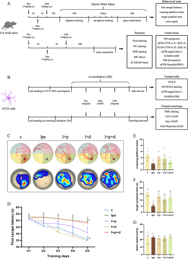
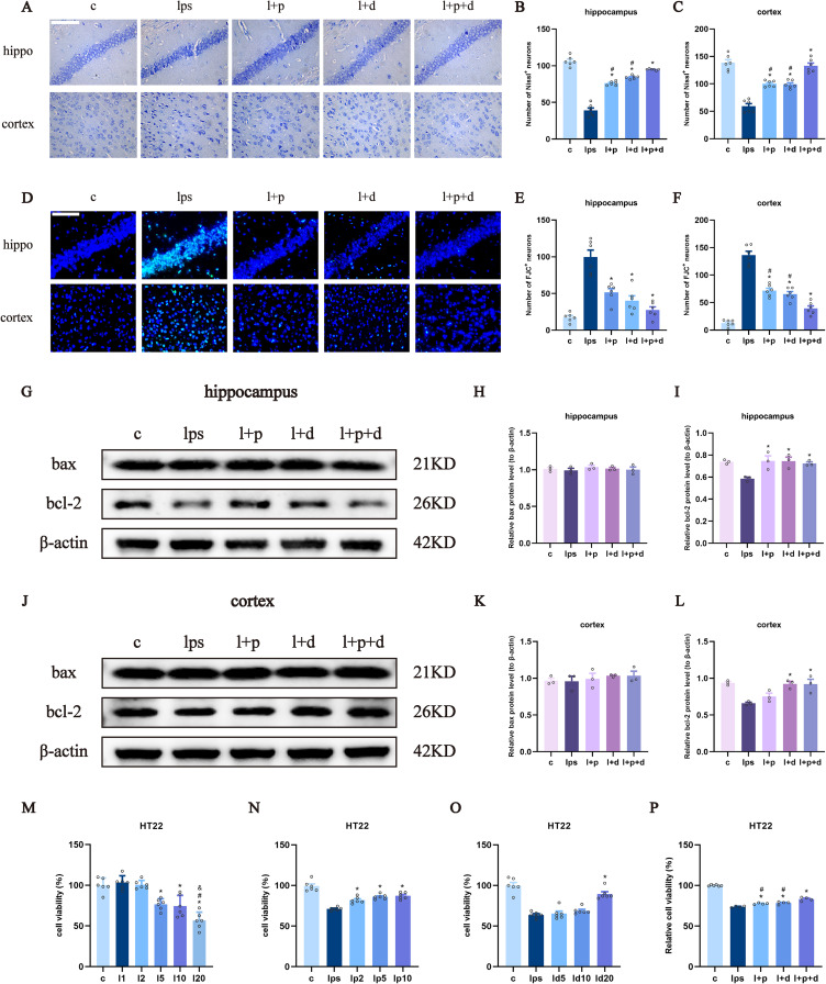
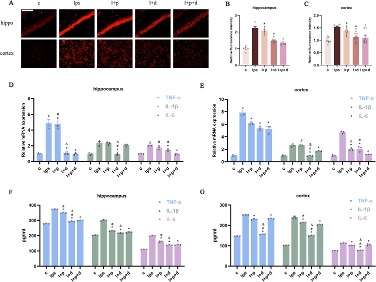
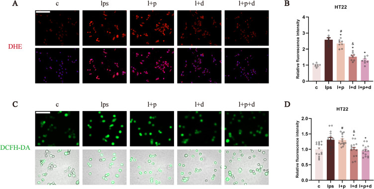
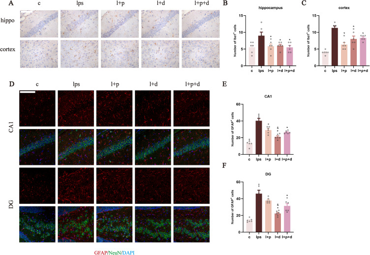
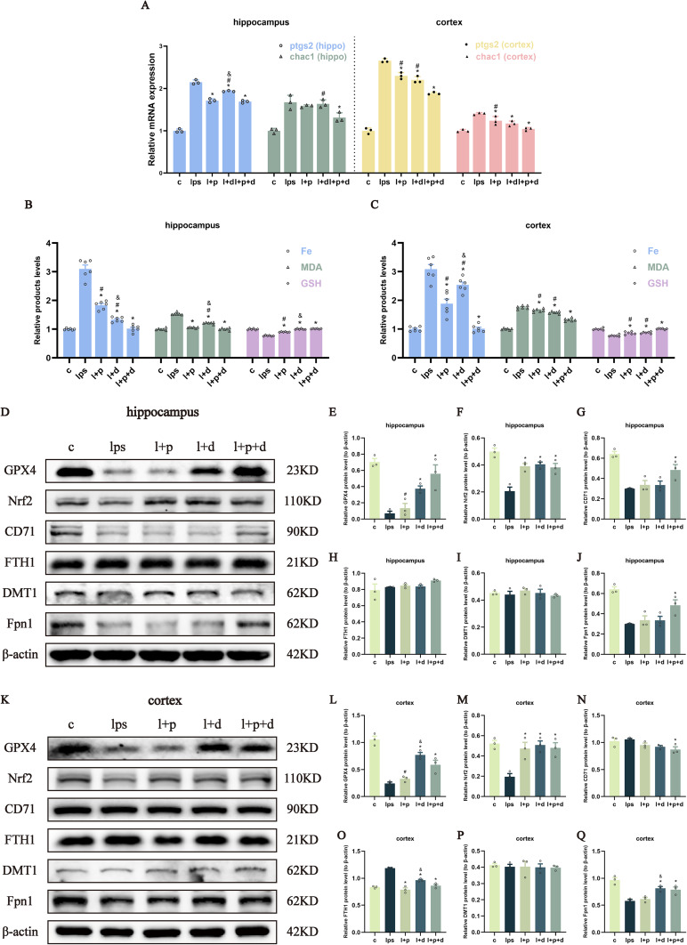
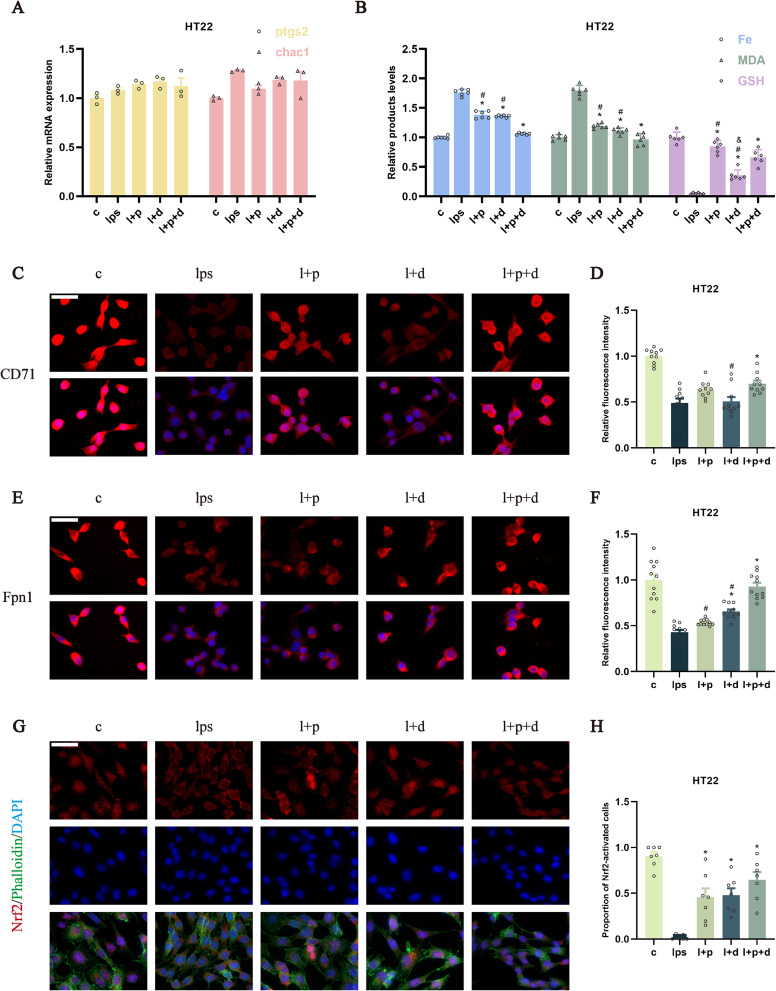
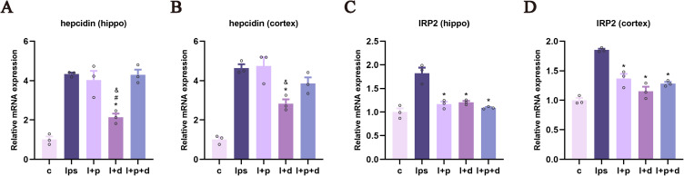
Methods: A total of 218 C57BL/6J male mice (20-25 g, 6-8 weeks) were used. Morris water maze (MWM) tests were performed to evaluate whether propofol and dexmedetomidine attenuated LPS-induced cognitive deficits. Brain injury was evaluated using Nissl and Fluoro-Jade C (FJC) staining. Neuroinflammation was assessed by dihydroethidium (DHE) and DCFH-DA staining and by measuring the levels of three cytokines. The number of Iba1+ and GFAP+ cells was used to detect the activation of microglia and astrocytes. To explore the involvement of ferroptosis, the levels of ptgs2 and chac1; the content of iron, malondialdehyde (MDA), and glutathione (GSH); and the expression of ferroptosis-related proteins were investigated.

Conclusion: The single use of propofol and dexmedetomidine mitigated LPS-induced cognitive impairment, while the combination showed poor performance. In alleviating endotoxemic neural loss and degeneration, the united sedative group exhibited the most potent capability. Both propofol and dexmedetomidine inhibited neuroinflammation, while propofol's effect was slightly weaker. All sedative groups reduced the neural apoptosis, inhibited the activation of microglia and astrocytes, and relieved neurologic ferroptosis. The combined group was most prominent in combating genetic and biochemical alterations of ferroptosis. Fpn1 may be at the core of endotoxemia-related ferroptosis activation.

Keywords: dexmedetomidine; encephalopathy; endotoxemia; ferroptosis; propofol.


Conflict of interest statement

The authors confirm that there are no conflicts of interest in this work.

Figures

Figure 1
Figure 1
Propofol and dexmedetomidine ameliorated the endotoxemia-associated cognitive impairment. (A and B) The intervention strategy and experimental procedure for endotoxemia model in vivo and in vitro. (C) Representative trajectory and heatmaps of the MWM. (D) The escape latency for the first platform discovery over five consecutive days. (E) The crossing-platform times on the final day after platform removal. (F) The duration of time spent in the quadrant where the platform was located on the final day. (G) The swim speed on the final day. Data are expressed as the mean ± SEM (*p<0.05 vs c group; #p<0.05 vs lps group; n = 7).
Figure 2
Figure 2
Propofol and dexmedetomidine mitigated the LPS-induced neuronal loss and degeneration as well as apoptosis. (A–C) Representative images of Nissl staining and the number of Nissl-positive cells (400×), scale bars = 50 μm. (DF) Representative images of FJC staining and number of FJC-positive cells (400×), scale bars = 50μm. (GL) The Western blot bands and quantitative analysis of bax and bcl-2. (MP) Cell viability of treated HT22 cells measured by CCK-8. Data are expressed as the mean ± SEM (*p<0.05 vs lps and l2 group; #p<0.05 vs l+p+d and l5 group; &p<0.05 vs l10 group; n = 6).
Figure 3
Figure 3
Propofol and dexmedetomidine diminished LPS-mediated neuroinflammation in endotoxemic mice. (AC) DHE staining representative images and analysis of relative fluorescence intensity (400×), scale bar = 50 μm. (D and E) The relative mRNA expression of TNF-α, IL-1β, and IL-6 measured by qPCR; (F and G) The content of TNF-α, IL-1β and IL-6 detected by ELISA. Data are expressed as the mean ± SEM (*p<0.05 vs lps group; #p<0.05 vs l+p+d group; &p<0.05 vs l+p group; n = 6).
Figure 4
Figure 4
Dexmedetomidine and combination abrogated the ROS production in LPS-incubated HT22 cells. (A and B) The relative fluorescence intensity of DHE staining to evaluate the level of superoxide anions (400×), scale bar = 50 μm. (C and D) The relative fluorescence intensity of DCFH-DA staining to investigate the production of H2O2 (400×), scale bar = 50 μm. Data are expressed as the mean ± SEM (*p<0.05 vs lps group; #p<0.05 vs l+p+d group; &p<0.05 vs l+p group; n = 6).
Figure 5
Figure 5
Propofol and dexmedetomidine reduced LPS-activated microglia and astrocytes. (AC) The presentative images and the number of Iba1-positive microglia (400×), scale bar = 50 μm. (DF) The presentative images and the number of GFAP-positive astrocytes (red) co-labelled with Neun-positive neurons (green) in the CA1 and DG of hippocampus (200×), scale bar = 100 μm. Data are expressed as the mean ± SEM (*p<0.05 vs lps group; &p<0.05 vs l+p group; n = 6).
Figure 6
Figure 6
Propofol and dexmedetomidine attenuated LPS-initiated ferroptosis in endotoxemic mice. (A) The relative mRNA expression of ptgs2 and chac1. (B and C) The relative products level of Fe, MDA and GSH. (DQ) The Western blot bands and quantitative analysis of GPX4, Nrf2, CD71, FTH1, DMT1 and Fpn1. Data are expressed as the mean ± SEM (*p<0.05 vs lps group; #p<0.05 vs l+p+d group; &p<0.05 vs l+p group; n = 6).
Figure 7
Figure 7
Propofol and dexmedetomidine inhibited LPS-induced ferroptosis in HT22 cells. (A) The relative mRNA expression of ptgs2 and chac1. (B) The relative products level of Fe, MDA and GSH. (CF) The representative images and relative fluorescence intensity of CD71 and Fpn1 (630×), scale bar = 50 μm. (G and H) The representative images and the proportion of activated Nrf2 cells (630×), scale bar = 50 μm. Data are expressed as the mean ± SEM (*p<0.05 vs lps group; #p<0.05 vs l+p+d group; n = 6).
Figure 8
Figure 8
Propofol and dexmedetomidine influenced the transcriptional level of hepcidin and IRP2 in endotoxemic mice. (A and B) The relative mRNA expression of hepcidin. (C and D) The relative mRNA expression of IRP2. Data are expressed as the mean ± SEM (*p<0.05 vs lps group; #p<0.05 vs l+p+d group; &p<0.05 vs l+p group; n = 3).
Figure 9
Figure 9
Mechanisms of propofol and dexmedetomidine in alleviating endotoxemia-induced encephalopathy. (A-C) The protective effects of propofol, dexmedetomidine and combined usage against LPS-mediated neural loss and degeneration, neural apoptosis and neuroinflammation. Blue triangles, green squares, and red polygons correspond to propofol, dexmedetomidine and combined groups. The asterisk (*) was used to indicate the relative strength of inhibition of the specific pathological conditions by each sedative group. (D) Three aspects of laboratory evidence to evaluate the extent of ferroptosis. (E) The iron metabolism, hepcidin-Fpn1 regulation, IRP/IRE axis, and Nrf2 translocation involved in the LPS-induced neural ferroptosis.

Similar articles

Cited by

References

    1. Singer M, Deutschman CS, Seymour CW, et al. The third international consensus definitions for sepsis and septic shock (Sepsis-3). JAMA. 2016;315(8):801–810. doi:10.1001/jama.2016.0287 - DOI - PMC - PubMed
    1. Hughes CG, Mailloux PT, Devlin JW, et al. Dexmedetomidine or propofol for sedation in mechanically ventilated adults with sepsis. N Engl J Med. 2021;384(15):1424–1436. doi:10.1056/NEJMoa2024922 - DOI - PMC - PubMed
    1. van der Poll T, Shankar-Hari M, Wiersinga WJ. The immunology of sepsis. Immunity. 2021;54(11):2450–2464. doi:10.1016/j.immuni.2021.10.012 - DOI - PubMed
    1. Rajendrakumar SK, Revuri V, Samidurai M, et al. Peroxidase-mimicking nanoassembly mitigates lipopolysaccharide-induced endotoxemia and cognitive damage in the brain by impeding inflammatory signaling in macrophages. Nano Lett. 2018;18(10):6417–6426. doi:10.1021/acs.nanolett.8b02785 - DOI - PubMed
    1. Xin Y, Tian M, Deng S, et al. The key drivers of brain injury by systemic inflammatory responses after sepsis: microglia and neuroinflammation. Molecular Neurobiology. 2022;60(3):1369–1390. doi:10.1007/s12035-022-03148-z - DOI - PMC - PubMed