Review
. 2024 Mar 27;16(3):e57014.
doi: 10.7759/cureus.57014. eCollection 2024 Mar.

A Comprehensive Review and Practical Guide of the Applications of Evoked Potentials in Neuroprognostication After Cardiac Arrest

Affiliations
Review

A Comprehensive Review and Practical Guide of the Applications of Evoked Potentials in Neuroprognostication After Cardiac Arrest

Eduard Portell Penadés et al. Cureus. .

Abstract

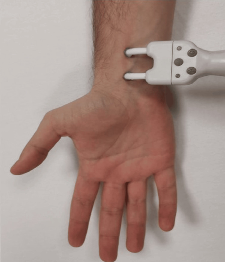
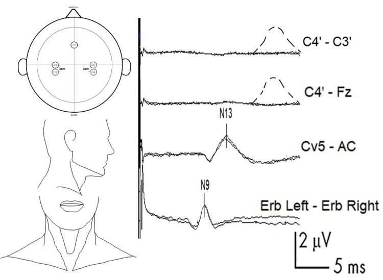
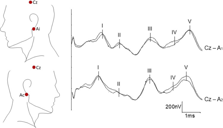
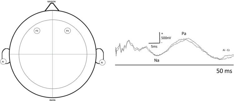
Cardiorespiratory arrest is a very common cause of morbidity and mortality nowadays, and many therapeutic strategies, such as induced coma or targeted temperature management, are used to reduce patient sequelae. However, these procedures can alter a patient's neurological status, making it difficult to obtain useful clinical information for the reliable estimation of neurological prognosis. Therefore, complementary investigations are conducted in the early stages after a cardiac arrest to clarify functional prognosis in comatose cardiac arrest survivors in the first few hours or days. Current practice relies on a multimodal approach, which shows its greatest potential in predicting poor functional prognosis, whereas the data and tools to identify patients with good functional prognosis remain relatively limited in comparison. Therefore, there is considerable interest in investigating alternative biological parameters and advanced imaging technique studies. Among these, somatosensory evoked potentials (SSEPs) remain one of the simplest and most reliable tools. In this article, we discuss the technical principles, advantages, limitations, and prognostic implications of SSEPs in detail. We will also review other types of evoked potentials that can provide useful information but are less commonly used in clinical practice (e.g., visual evoked potentials; short-, medium-, and long-latency auditory evoked potentials; and event-related evoked potentials, such as mismatch negativity or P300).

Keywords: brainstem auditory evoked potentials; event-related potentials; middle latency auditory evoked potentials; mismatch negativity; somatosensory evoked potentials; technical aspects.


Conflict of interest statement

The authors have declared that no competing interests exist.

Figures

Figure 1
Figure 1. Stimulation of the median nerve at the wrist level
The anode (positive electrode) should be at the distal position. Alternatively, subdermal needle electrodes can be used. Movement of the thumb should be confirmed to ensure sufficient intensity of stimulation (while being careful with curare).
Figure 2
Figure 2. Example of SSEP with absent N20 response and recommended electrode placement and normal curves
First channel (bottom line): N9 (normal value: approximately 9 ms) is generated in the trunks of the brachial plexus. This wave is recorded in the ipsilateral Erb-contralateral Erb channel. The Erb point is located within the angle formed by the posterior border of the clavicular head of the sternocleidomastoid muscle and the clavicle, 2-3 cm posterior to the clavicle. Second channel (middle line): N13 (normal value: approximately 13 ms) is generated by the postsynaptic activity of the dorsal horn of the cervical spinal cord. This wave is recorded from Cv5 (placed on the skin over the fifth cervical spine), and the AC (placed in the anterior cervical region) is used as the reference. Top channel: example of an absent N20. The dashed line represents the expected curve, which was generated in the primary sensitive cortex of the postcentral gyrus (normal value: approximately 20 ms). This wave is recorded from C3' and C4' (2-3 cm behind C3 and C4) (contralateral side as the active electrode, ipsilateral side as the reference) or by using Fz as the reference instead. SSEP: somatosensory evoked potential
Figure 3
Figure 3. BAEPs: recommended electrode placement and normal curves
The reference for both channels will be Cz (the skull), with the active electrode on the ipsilateral earlobe (Ai) in one channel and on the contralateral earlobe (Ac) in the other channel. The mastoid can be used instead of the ear (called in this case Mi: ipsilateral or Mc: contralateral). The five main waves are named sequentially using Roman numerals in the order of appearance from peripheral to central (I, II, III, IV, and V). BAEPs: brainstem auditory evoked potentials
Figure 4
Figure 4. MLAEPs: recommended electrode placement and normal curves
Two channels with the active electrode located over both primary auditory cortices (F3 and F4) and with a common reference to the ipsilateral earlobe or mastoid. A negative wave called Na, of debated origin but probably thalamic, appears at approximately 17 ms after the stimulus, and a second positive wave at around 30 ms after the stimulus, called Pa, is generated in the primary auditory cortex. MLAEPs: middle-latency auditory evoked potentials Courtesy of Dr. Misericordia Veciana and Dr. Jordi Pedro Pérez, Neurophysiology Department, Hospital Universitari de Bellvitge, Barcelona, Spain
Figure 5
Figure 5. MMN curve
Figure 5 shows two examples: on top, a patient with an MMN present, and on the bottom, a patient with MMN absent. We observe the averages after a series of standard stimuli (s) and the responses after rare stimuli (r). Subtracting the standard from the rare averages yielded the MMN. The first large negative wave observed at approximately 100 ms is called N100 or N1 and is generated in the auditory cortex, the presence of which is required to obtain MMN. The electrodes are placed according to the international 10-20 system, with the active electrode in the midline (Fz, Cz, or Pz), preferably in Fz, and with reference in the mastoids or earlobes, linked or not linked. MMN: mismatch negativity Courtesy of Dr. Misericordia Veciana and Dr. Jordi Pedro Pérez, Neurophysiology Department, Hospital Universitari de Bellvitge, Barcelona, Spain

Similar articles

References

    1. Heart Disease and Stroke Statistics-2022 update: a report from the American Heart Association. Tsao CW, Aday AW, Almarzooq ZI, et al. Circulation. 2022;145:0–639. - PubMed
    1. Global incidences of out-of-hospital cardiac arrest and survival rates: systematic review of 67 prospective studies. Berdowski J, Berg RA, Tijssen JG, Koster RW. Resuscitation. 2010;81:1479–1487. - PubMed
    1. Heart Disease and Stroke Statistics-2020 update: a report from the American Heart Association. Virani SS, Alonso A, Benjamin EJ, et al. Circulation. 2020;141:0–596. - PubMed
    1. EuReCa ONE-27 Nations, ONE Europe, ONE Registry: a prospective one month analysis of out-of-hospital cardiac arrest outcomes in 27 countries in Europe. Gräsner JT, Lefering R, Koster RW, et al. Resuscitation. 2016;105:188–195. - PubMed
    1. Brain injury after cardiac arrest. Perkins GD, Callaway CW, Haywood K, et al. Lancet. 2021;398:1269–1278. - PubMed

LinkOut - more resources