

Prevalence and Variations of the Median Artery: A Pilot Study in a Sample of Lithuanian Cadavers
- PMID: 38681388
- PMCID: PMC11055626
- DOI: 10.7759/cureus.57140
Prevalence and Variations of the Median Artery: A Pilot Study in a Sample of Lithuanian Cadavers
Abstract
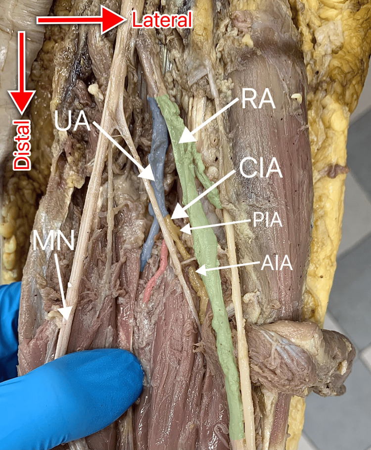
Objective This pilot project aimed to assess the prevalence and variations of the median artery (MA) on a small scale in preparation for a large-scale study investigating MA in Lithuanian cadavers. Methods Eight formalin-fixed adult female cadavers were used in this study. Dissection was performed to allow for the observation of MA presence, type, origin, termination, and relations with other structures. The gathered data was analyzed, and a literature search was performed to compare the findings. Results MA was found in 10 of the 16 upper limbs examined; therefore, the incidence of MA in the present study was 62.5%. Of the 10 MAs found, six (60%) were of the antebrachial type (a-MA), and four (40%) were palmar (p-MA). Thus, the prevalence of a-MA and p-MA in the upper limbs examined was 37.5% (N = 6/16) and 25% (N = 4/16), respectively. Among the six cadavers that were found to possess MA, it was identified bilaterally in four (66.7%) and unilaterally in two (33.3%). The associations between the antimere and the presence of MA or MA-type were not statistically significant. MA most commonly originated from the common interosseous artery (50%, N = 5/10), followed by the ulnar artery (UA) (40%, N = 4/10), and the anterior interosseous artery (10%, N = 1/10). Two (33.3%) of the six a-MAs terminated in the mid-forearm, while four (66.7%) a-MAs ended in the distal forearm. Meanwhile, three (75%) of the four p-MAs terminated by joining the UA, while one (25%) terminated as the first common palmar digital artery. In the forearm, nine (90%) of the 10 MAs traveled anteriorly to the anterior interosseous nerve (AIN), and only one (10%) traveled posteriorly to the AIN. Additionally, one (10%) of the 10 MAs was found to pierce the median nerve. Conclusions Our findings confirm the variability in MA characteristics reported by previous studies. The high incidence of MA discovered in our sample calls attention to the importance of being aware of MA in a clinical setting, as this would allow for a timely and accurate response to a potential pathology associated with this structure.
Keywords: anatomical variations; antebrachial type; arteries of the upper limb; cadaveric study; median artery; palmar type; persistent median artery.
Copyright © 2024, Berškys et al.
Conflict of interest statement
The authors have declared that no competing interests exist.
Figures


Similar articles
-
Median artery revisited.J Anat. 1999 Jul;195 ( Pt 1)(Pt 1):57-63. doi: 10.1046/j.1469-7580.1999.19510057.x. J Anat. 1999. PMID: 10473293 Free PMC article.
-
A preliminary survey of the median artery in human cadavers of South Indian origin.Bratisl Lek Listy. 2011;112(5):292-5. Bratisl Lek Listy. 2011. PMID: 21682087
-
An Anatomical Variation of Median Artery of Forearm and Palm: Cadaveric Study into its Origin and Course.Kathmandu Univ Med J (KUMJ). 2022 Oct-Dec;20(80):443-447. Kathmandu Univ Med J (KUMJ). 2022. PMID: 37795721
-
Median artery of the forearm in human fetuses in northeastern Brazil: anatomical study and review of the literature.Anat Sci Int. 2017 Jan;92(1):107-111. doi: 10.1007/s12565-015-0322-x. Epub 2016 Jan 8. Anat Sci Int. 2017. PMID: 26747631 Review.
-
High origin of a superficial ulnar artery arising from the axillary artery: anatomy, embryology, clinical significance and a review of the literature.Folia Morphol (Warsz). 2006 Nov;65(4):400-5. Folia Morphol (Warsz). 2006. PMID: 17171623 Review.
References
-
- Persistent median artery as a cause of pronator syndrome. Jones NF, Ming NL. J Hand Surg Am. 1988;13:728–732. - PubMed
-
- Arterial patterns of the human upper limb: update of anatomical variations and embryological development. Rodríguez-Niedenführ M, Vázquez T, Parkin IG, Sañudo JR. http://www.eurjanat.com/web/paper.php?id=03S10021 Eur J Anat. 2003;7:21–28.
-
- Persistent median artery in the carpal tunnel: anatomy, embryology, clinical significance, and review of the literature. Natsis K, Iordache G, Gigis I, et al. https://journals.viamedica.pl/folia_morphologica/article/view/15898. Folia Morphol (Warsz) 2009;68:193–200. - PubMed
-
- Median artery of the forearm in human fetuses in northeastern Brazil: anatomical study and review of the literature. Aragão JA, da Silva AC, Anunciação CB, Reis FP. Anat Sci Int. 2017;92:107–111. - PubMed
LinkOut - more resources
Full Text Sources