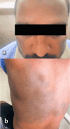
Bilateral Naevus of Ito and Ota With Palatal Involvement
- PMID: 38681431
- PMCID: PMC11046169
- DOI: 10.7759/cureus.57004
Bilateral Naevus of Ito and Ota With Palatal Involvement
Abstract
Naevus of Ito and naevus of Ota are benign dermal melanocytoses with similar pathogenic mechanisms of failure in the melanocyte migration to typical locations within the basal layer from neural crest cells and differ in distribution. Bilateral and oral mucosal involvement of naevus of Ota can occur but is infrequent. Naevus of Ito is seldom associated with naevus of Ota and extracutaneous manifestations. A review of the English literature showed 14 cases of naevus of Ota with palatal involvement. None showed bilateral involvement of both naevi with oral involvement. Here we report the case of bilateral naevus of Ito and bilateral naevus of Ota with palatal involvement. A 32-year-old male came to us with naevus of Ito on both sides of his back and naevus of Ota on both sides of his face involving the sclera of both eyes with a bluish lesion along the midline of the hard palate.
Keywords: acquired nevi; congenital melanocytic nevi; hori naevus; naevus of ito; naevus of ota.
Copyright © 2024, Pillai et al.
Conflict of interest statement
The authors have declared that no competing interests exist.
Figures


Similar articles
-
Unilateral Nevus of Ota with Bilateral Nevus of Ito and Palatal Lesion: A Case Report with a Proposed Clinical Modification of Tanino's Classification.Indian J Dermatol. 2013 Jul;58(4):286-9. doi: 10.4103/0019-5154.113943. Indian J Dermatol. 2013. PMID: 23918999 Free PMC article.
-
Case report: nevus of Ota and nevus of Ito associated with meningeal melanocytosis.Neurocirugia (Engl Ed). 2020 Nov-Dec;31(6):299-305. doi: 10.1016/j.neucir.2019.10.001. Epub 2019 Nov 25. Neurocirugia (Engl Ed). 2020. PMID: 31780112 English, Spanish.
-
Dermal dendritic melanocytic proliferations: an update.Histopathology. 2004 Nov;45(5):433-51. doi: 10.1111/j.1365-2559.2004.01975.x. Histopathology. 2004. PMID: 15500647 Review.
-
Bilateral naevus of Ota: a rare manifestation in a Caucasian.J Eur Acad Dermatol Venereol. 2004 May;18(3):353-5. doi: 10.1111/j.1468-3083.2004.00857.x. J Eur Acad Dermatol Venereol. 2004. PMID: 15096155
-
[Treatment of nevus of Ota and Ito and epidermal nevus syndrome].Hautarzt. 2020 Dec;71(12):926-931. doi: 10.1007/s00105-020-04710-3. Hautarzt. 2020. PMID: 33145623 Review. German.
References
-
- Bilateral nevus of Ota with palatal involvement, unilateral nevus of Ito, and a port-wine stain: a rare presentation. Mohta A, Kushwaha RK, Kumari P, Shamra MK, Jain SK. Our Dermatol Online. 2022;13:197–201.
-
- Natural history of nevus of Ota. Hidano A, Kajima H, Ikeda S, Mizutani H, Miyasato H, Niimura M. https://pubmed.ncbi.nlm.nih.gov/6018994/ Arch Dermatol. 1967;95:187–195. - PubMed
-
- Nevus of Ota with buccal mucosal pigmentation: a rare case. Shetty SR, Subhas BG, Rao KA, Castellino R. https://www.ncbi.nlm.nih.gov/pmc/articles/PMC3177382/ Dent Res J (Isfahan) 2011;8:52–55. - PMC - PubMed
-
- Dermal dendritic melanocytic proliferations: an update. Zembowicz A, Mihm MC. Histopathology. 2004;45:433–451. - PubMed
-
- The naevus of Ota. Swann PG, Kwong E. Clin Exp Optom. 2010;93:264–267. - PubMed
Publication types
LinkOut - more resources
Full Text Sources
