

Vascular Malformation Involving the Brachial Plexus: A Case Report and Review of Literature
- PMID: 38681915
- PMCID: PMC11043966
- DOI: 10.13107/jocr.2024.v14.i04.4372
Vascular Malformation Involving the Brachial Plexus: A Case Report and Review of Literature
Abstract
Introduction: Vascular anomalies, comprising up to 4.5% of the general population, are aberrations occurring during vascular development. Vascular abnormalities are frequently identified in children and frequently exhibit characteristics similar to nerve sheath tumors. We report a case of 16 years old boy with a arterio-venous (AV) malformation (AVM) affecting the brachial plexus. We discuss the clinical features, diagnosis, treatment, and histopathological findings in this patient and review the relevant literature.
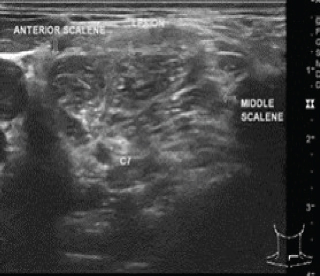
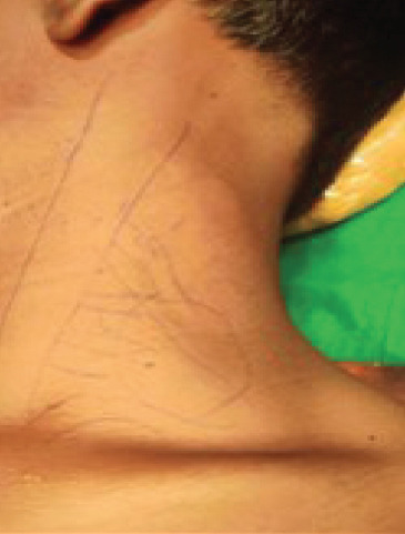
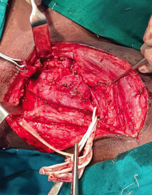
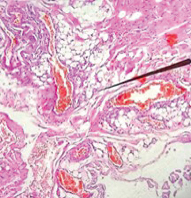
Case report: A 16- year-s old boy presented with pain, paresthesia, swelling, and reduced grip strength of the hand. Radiological investigations revealed a vascular lesion encasing C5, C6 nerve roots and displacing the C7 root. Near total surgical excision of the lesion was done with preservation of nerve. Histopathology confirmed arteriovenous AVMmalformation with distinct features.
Conclusion: High-resolution ultrasound is crucial for diagnosing soft- tissue vascular anomalies. Surgeons well versed in micro surgical skill play a vital key role in minimizing neural deficits. In the case of vascular malformations of brachial plexus, near total excision is the most favorable option.
Keywords: Vascular anomalies; arterio-venous malformation (AVM); brachial plexus.
Copyright: © Indian Orthopaedic Research Group.
Conflict of interest statement
Conflict of Interest: Nil
Figures
Similar articles
-
The C5 root dermatome enlarges and modulates hand pain in total brachial plexus palsy.Microsurgery. 2014 May;34(4):292-5. doi: 10.1002/micr.22210. Microsurgery. 2014. PMID: 24822255
-
Contralateral C7 transfers: An innovative approach to improving peripheral neuropathic pain after traumatic brachial plexus injury with C5 rupture and avulsion of C6, C7, C8 and T1. A case series study.Clin Neurol Neurosurg. 2020 Apr;191:105693. doi: 10.1016/j.clineuro.2020.105693. Epub 2020 Jan 23. Clin Neurol Neurosurg. 2020. PMID: 32035358
-
Management of Adult Traumatic Brachial Plexus Injury.Mymensingh Med J. 2023 Apr;32(2):437-447. Mymensingh Med J. 2023. PMID: 37002755
-
Brachial plexus neuropathy by hibernoma compression: An unusual presentation of peripheral non-neural sheath nerve tumor and a systematic review of literature.Neurol India. 2019 Mar-Apr;67(2):481-484. doi: 10.4103/0028-3886.258049. Neurol India. 2019. PMID: 31085864
-
Venous malformation serving as the draining vein of an adjoining arteriovenous malformation. Case report and review of the literature.Surg Neurol. 2001 Sep;56(3):170-4. doi: 10.1016/s0090-3019(01)00457-8. Surg Neurol. 2001. PMID: 11597644 Review.
References
-
- Greene AK. Vascular anomalies:Current overview of the field. Clin Plast Surg. 2011;38:1–5. - PubMed
-
- Rusu GM, Ciuce C, Fodor L, Manole S, Dudea SM. Ultrasonographic and imaging appearance of peripheral intraneural vascular anomalies:Report of two cases and review of the literature. Med Ultrason. 2018;20:237–46. - PubMed
-
- Donnelly LF, Adams DM, Bisset GS., 3rd Vascular malformations and hemangiomas:A practical approach in a multidisciplinary clinic. Am J Roentgenol. 2000;174:597608. - PubMed
-
- McCormack HM, Horne DJ, Sheather S. Clinical applications of visual analogue scales:A critical review. Psychol Med. 1988;18:1007–19. - PubMed
-
- Mulliken JB, Glowacki J. Hemangiomas and vascular malformations in infants and children:A classification based on endothelial characteristics. Plast Reconstr Surg. 1982;69:412–22. - PubMed
Publication types
LinkOut - more resources
Full Text Sources
Miscellaneous