Sagittal balance parameters measurement on cervical spine MR images based on superpixel segmentation
- PMID: 38681963
- PMCID: PMC11048045
- DOI: 10.3389/fbioe.2024.1337808
Sagittal balance parameters measurement on cervical spine MR images based on superpixel segmentation
Abstract
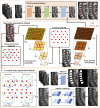
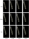
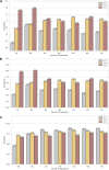
Introduction: Magnetic Resonance Imaging (MRI) is essential in diagnosing cervical spondylosis, providing detailed visualization of osseous and soft tissue structures in the cervical spine. However, manual measurements hinder the assessment of cervical spine sagittal balance, leading to time-consuming and error-prone processes. This study presents the Pyramid DBSCAN Simple Linear Iterative Cluster (PDB-SLIC), an automated segmentation algorithm for vertebral bodies in T2-weighted MR images, aiming to streamline sagittal balance assessment for spinal surgeons. Method: PDB-SLIC combines the SLIC superpixel segmentation algorithm with DBSCAN clustering and underwent rigorous testing using an extensive dataset of T2-weighted mid-sagittal MR images from 4,258 patients across ten hospitals in China. The efficacy of PDB-SLIC was compared against other algorithms and networks in terms of superpixel segmentation quality and vertebral body segmentation accuracy. Validation included a comparative analysis of manual and automated measurements of cervical sagittal parameters and scrutiny of PDB-SLIC's measurement stability across diverse hospital settings and MR scanning machines. Result: PDB-SLIC outperforms other algorithms in vertebral body segmentation quality, with high accuracy, recall, and Jaccard index. Minimal error deviation was observed compared to manual measurements, with correlation coefficients exceeding 95%. PDB-SLIC demonstrated commendable performance in processing cervical spine T2-weighted MR images from various hospital settings, MRI machines, and patient demographics. Discussion: The PDB-SLIC algorithm emerges as an accurate, objective, and efficient tool for evaluating cervical spine sagittal balance, providing valuable assistance to spinal surgeons in preoperative assessment, surgical strategy formulation, and prognostic inference. Additionally, it facilitates comprehensive measurement of sagittal balance parameters across diverse patient cohorts, contributing to the establishment of normative standards for cervical spine MR imaging.
Keywords: artificial intelligence; cervical spine; magnetic resonance imaging; sagittal balance parameters; superpixel segmentation.
Copyright © 2024 Zhong, Dai, Li, Zhu, Lin, Ran, Chen, Ruan, Yu, Li, Li, Xu, Sun, Weber, Kong, Yang, Lin, Chen, Chen, Jiang, Zhou, Sheng, Wang, Tian and Sun.
Conflict of interest statement
The authors declare that the research was conducted in the absence of any commercial or financial relationships that could be construed as a potential conflict of interest.
Figures


Similar articles
-
Image Segmentation of Brain MRI Based on LTriDP and Superpixels of Improved SLIC.Brain Sci. 2020 Feb 20;10(2):116. doi: 10.3390/brainsci10020116. Brain Sci. 2020. PMID: 32093401 Free PMC article.
-
Automatic cell segmentation in histopathological images via two-staged superpixel-based algorithms.Med Biol Eng Comput. 2019 Mar;57(3):653-665. doi: 10.1007/s11517-018-1906-0. Epub 2018 Oct 16. Med Biol Eng Comput. 2019. PMID: 30327998
-
Predicting the risk and severity of acute spinal cord injury after a minor trauma to the cervical spine.Spine J. 2013 Jun;13(6):597-604. doi: 10.1016/j.spinee.2013.02.006. Epub 2013 Mar 21. Spine J. 2013. PMID: 23523437
-
Optimized method for segmentation of ancient mural images based on superpixel algorithm.Front Neurosci. 2022 Nov 2;16:1031524. doi: 10.3389/fnins.2022.1031524. eCollection 2022. Front Neurosci. 2022. PMID: 36408409 Free PMC article.
-
Semi-Automatic Segmentation of Vertebral Bodies in MR Images of Human Lumbar Spines.Appl Sci (Basel). 2018 Sep;8(9):1586. doi: 10.3390/app8091586. Epub 2018 Sep 7. Appl Sci (Basel). 2018. PMID: 30637136 Free PMC article.
References
-
- Amin A., Abbas M., Salam A. A. (2022). “Automatic detection and classification of scoliosis from spine X-rays using transfer learning,” in 2nd International Conference on Digital Futures and Transformative Technologies (ICoDT2),2022, 1–6. 10.1109/ICoDT255437 - DOI
-
- Barbieri P. D., Pedrosa G. V., Traina A. J. M., Nogueira-Barbosa M. H.(2015). Vertebral body segmentation of spine MR images using superpixels, IEEE 28th Int. Symposium Computer-Based Med. Syst., 44–49. 10.1109/CBMS.2015.11 - DOI
-
- Boudreau C., Carrondo Cottin S., Ruel-Laliberté J., Mercier D., Gélinas-Phaneuf N., Paquet J. (2021). Correlation of supine MRI and standing radiographs for cervical sagittal balance in myelopathy patients: a cross-sectional study. Eur. Spine J. 30, 1521–1528. 10.1007/s00586-021-06833-0 - DOI - PubMed
Grants and funding
LinkOut - more resources
Full Text Sources

