. 2024 May 15;151(10):dev202475.
doi: 10.1242/dev.202475. Epub 2024 May 17.

Optical coherence tomography-guided Brillouin microscopy highlights regional tissue stiffness differences during anterior neural tube closure in the Mthfd1l murine mutant

Affiliations

Optical coherence tomography-guided Brillouin microscopy highlights regional tissue stiffness differences during anterior neural tube closure in the Mthfd1l murine mutant

Yogeshwari S Ambekar et al. Development. .

Abstract

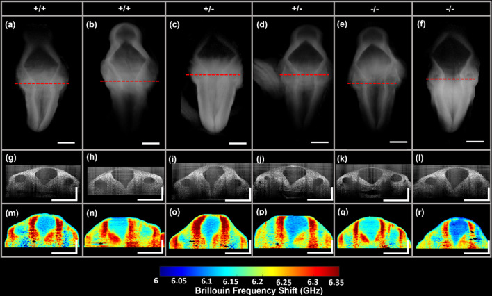
Neurulation is a highly synchronized biomechanical process leading to the formation of the brain and spinal cord, and its failure leads to neural tube defects (NTDs). Although we are rapidly learning the genetic mechanisms underlying NTDs, the biomechanical aspects are largely unknown. To understand the correlation between NTDs and tissue stiffness during neural tube closure (NTC), we imaged an NTD murine model using optical coherence tomography (OCT), Brillouin microscopy and confocal fluorescence microscopy. Here, we associate structural information from OCT with local stiffness from the Brillouin signal of embryos undergoing neurulation. The stiffness of neuroepithelial tissues in Mthfd1l null embryos was significantly lower than that of wild-type embryos. Additionally, exogenous formate supplementation improved tissue stiffness and gross embryonic morphology in nullizygous and heterozygous embryos. Our results demonstrate the significance of proper tissue stiffness in normal NTC and pave the way for future studies on the mechanobiology of normal and abnormal embryonic development.

Keywords: Biomechanics; Brillouin microscopy; Mouse; Neural tube closure; Neural tube defects.


Conflict of interest statement

Competing interests M.S. and K.V.L. have a financial interest in ElastEye, which is unrelated to this work. All other authors declare they have no competing interests.

Figures

Fig. 1.
Fig. 1.
The one-carbon metabolism pathway is required for neurulation. Neural tube defects are a highly penetrant phenotype in the Mthfd1l knockout mice lineage. Failures during neural fold closure are identified by immunostainings against Pax6, represented in white in the top images. The neuron-specific β-tubulin (Tubb3) is shown in red. The diagram represents the compartmentalization of the one-carbon metabolism between the cytosol and mitochondria. The gene Mthfd1l encodes a mitochondrial monofunctional enzyme responsible for catalyzing 10-formyl-tetrahydrofolate to formate, which is the last step in the flow of one-carbon units from the mitochondria to the cytoplasm.
Fig. 2.
Fig. 2.
Mthfd1l ablation decreases tissue stiffness in E9.5 embryos. (A-G) 3D-OCT images showing the hindbrains of Mthfd1l embryos at E9.5. (H-N) 2D-OCT optical sections showing the neural folds at rhombomere 5 near the otic pits. (O-U) Brillouin frequency shift images represent tissue stiffness in the anatomical areas identified by the corresponding 2D-OCT optical sections. The stiffness measurements by OCT-Brillouin were repeated at least four times per genotype. Red dashed line indicates the plane used for the respective (H-N) 2D OCT and (O-U) Brillouin imaging. Yellow arrows indicate abnormal neural tube phenotypes. Scale bars: 0.25 mm.
Fig. 3.
Fig. 3.
Mthfd1l ablation decreases tissue stiffness in E10.5 embryos. (A-F) 3D-OCT images showing the hindbrains of Mthfd1l embryos at E10.5. (G-L) 2D-OCT optical sections showing the neural folds at rhombomere 5 near the otic pits. (M-R) Brillouin frequency shift images represent tissue stiffness in the anatomical areas identified by the corresponding 2D-OCT optical sections. The stiffness measurements by OCT-Brillouin were repeated at least four times per genotype. Red dashed line indicates the plane used for the respective (H-N) 2D OCT and (O-U) Brillouin imaging. Scale bars: 0.5 mm.
Fig. 4.
Fig. 4.
Formate supplementation improves tissue stiffness in E9.5 embryos. (A-F) 3D-OCT images showing the hindbrains of formate-supplemented Mthfd1l embryos at E9.5. (G-L) 2D-OCT optical sections showing the neural folds at rhombomere 5 near the otic pits. (M-R) Brillouin frequency shift images represent tissue stiffness in the anatomical areas identified by the corresponding 2D-OCT optical sections. The stiffness measurements by OCT-Brillouin were repeated at least four times per genotype. Red dashed line indicates the plane used for the respective (H-N) 2D OCT and (O-U) Brillouin imaging. Scale bars: 0.25 mm.
Fig. 5.
Fig. 5.
Formate supplementation improves tissue stiffness in E10.5 embryos. (A-F) 3D-OCT images showing the hindbrains of formate-supplemented Mthfd1l embryos at E10.5. (G-L) 2D-OCT optical sections showing the neural folds at rhombomere 5 near the otic pits. (M-R) Brillouin frequency shift images represent tissue stiffness in the anatomical areas identified by the corresponding 2D-OCT optical sections. The stiffness measurements by OCT-Brillouin were repeated at least four times per genotype. Red dashed line indicates the plane used for the respective (H-N) 2D OCT and (O-U) Brillouin imaging. Scale bars: 0.5 mm.
Fig. 6.
Fig. 6.
Region-wise average Brillouin frequency shift of embryos. (A-D) Region-wise average Brillouin frequency shift of wild-type (WT), heterozygous (HET) and nullizygous (NULL) embryos at the neural tube neuroepithelia (A), adjacent paraxial mesenchyme (B), otic pit (C) and non-neural surface ectoderm region (D), without supplementation (No Supp.) and with formate supplementation (Supp.) at E9.5 and E10.5. The data are represented as the inter-sample mean±s.d., and the mean per sample are plotted alongside as respective individual points. *P<0.05, **P<0.01, ***P<0.001 (Kruskal–Wallis ANOVA and pairwise Dunn's test).
Fig. 7.
Fig. 7.
Mthfd1l ablation decreases neuronal differentiation. (A-R) Representative images showing whole mount Pax6 immunostaining (A-C,J-L), whole mount Tubb3 immunostaining (D-F,M-O) and Pax6 and Tubb3 merged channels of fluorescence imaging (G-I,P-R) at the level of the first and second pharyngeal arches. Pax6-positive neural progenitor cells are decreased in Mthfd1l mutant embryos, as indicated by the arrows in B and C. The arrows in E and F indicate decreased neural differentiation detected along the pharyngeal arches using the Tubb3 antibody, which indicates a neuron-specific β-tubulin. Formate supplementation (J-R) re-establishes Pax6-positive progenitor cells and respective neural differentiation. Mthfd1l embryos were immunostained against Pax6 and Tubb3 at E10.0. The immunostaining and imaging were repeated at least three times per genotype. Scale bars: 0.5 mm.
Fig. 8.
Fig. 8.
Mthfd1l ablation disrupts neural crest cell migration and somitogenesis. (A-L) Representative images showing immunostaining of Mthfd1l embryos at E9.5; whole mount Sox10 immunostaining (A-C,G-I), whole mount Pax3 immunostaining (D-F,J-L). Sox 10-positive neural crest cells are detected in decreased levels in Mthfd1l mutant embryos, as indicated by the arrows in B and C. Pax3 staining of somites indicates abnormal somitogenesis, as indicated by the arrows in E and F. Formate supplementation restores the levels of Sox 10 and Pax3 (G-L). The immunostaining and imaging were repeated at least three times per genotype. Scale bars: 0.5 mm.

Similar articles

References

    1. Ambekar, Y. S., Singh, M., Zhang, J., Nair, A., Aglyamov, S. R., Scarcelli, G. and Larin, K. V. (2020). Multimodal quantitative optical elastography of the crystalline lens with optical coherence elastography and Brillouin microscopy. Biomed. Opt. Express 11, 2041-2051. 10.1364/BOE.387361 - DOI - PMC - PubMed
    1. Ambekar, Y. S., Singh, M., Schill, A. W., Zhang, J., Zevallos-Delgado, C., Khajavi, B., Aglyamov, S. R., Finnell, R. H., Scarcelli, G. and Larin, K. V. (2022). Multimodal imaging system combining optical coherence tomography and Brillouin microscopy for neural tube imaging. Opt. Lett. 47, 1347-1350. 10.1364/OL.453996 - DOI - PMC - PubMed
    1. Armistead, J., Patel, N., Wu, X., Hemming, R., Chowdhury, B., Basra, G. S., Del Bigio, M. R., Ding, H. and Triggs-Raine, B. (2015). Growth arrest in the ribosomopathy, Bowen-Conradi syndrome, is due to dramatically reduced cell proliferation and a defect in mitotic progression. Biochim. Biophys. Acta 1852, 1029-1037. 10.1016/j.bbadis.2015.02.007 - DOI - PubMed
    1. Barriga, E. H., Franze, K., Charras, G. and Mayor, R. (2018). Tissue stiffening coordinates morphogenesis by triggering collective cell migration in vivo. Nature 554, 523-527. 10.1038/nature25742 - DOI - PMC - PubMed
    1. Bischof, C., Mirtschink, P., Yuan, T., Wu, M., Zhu, C., Kaur, J., Pham, M. D., Gonzalez-Gonoggia, S., Hammer, M., Rogg, E.-M.et al. (2021). Mitochondrial-cell cycle cross-talk drives endoreplication in heart disease. Sci. Transl. Med. 13, eabi7964. 10.1126/scitranslmed.abi7964 - DOI - PubMed

MeSH terms

Substances