

Cutibacterium acnes-derived extracellular vesicles promote tumor growth in renal cell carcinoma
- PMID: 38682309
- PMCID: PMC11309925
- DOI: 10.1111/cas.16202
Cutibacterium acnes-derived extracellular vesicles promote tumor growth in renal cell carcinoma
Abstract
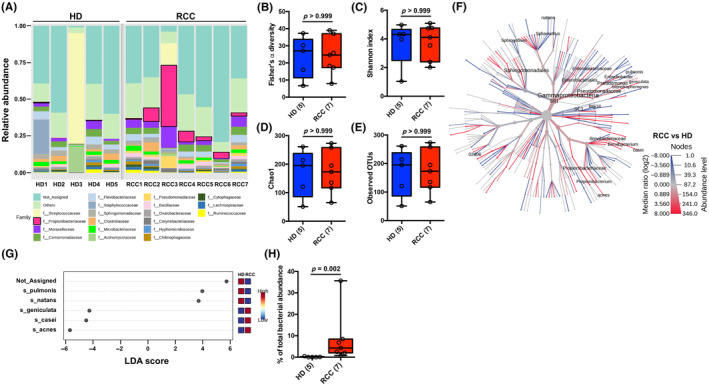
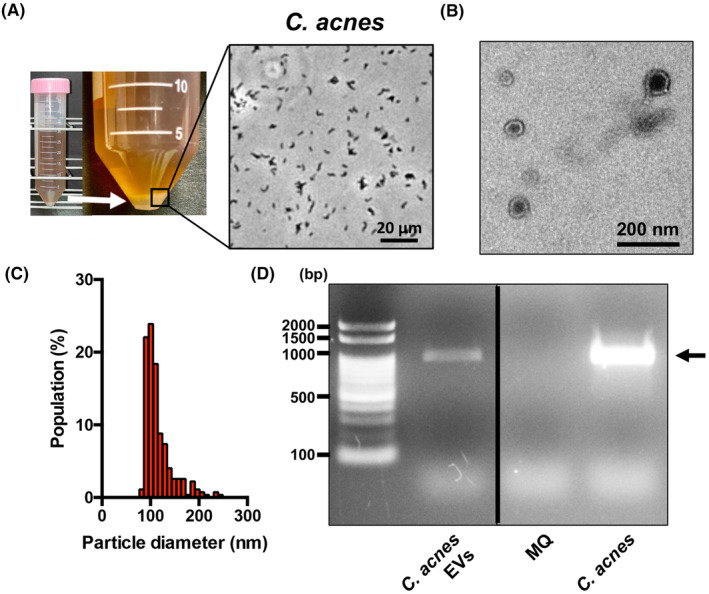
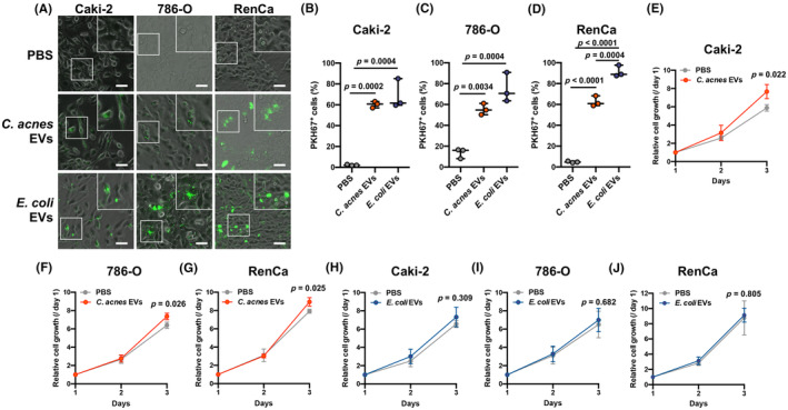
Bacterial flora are present in various parts of the human body, including the intestine, and are thought to be involved in the etiology of various diseases such as multiple sclerosis, intestinal diseases, cancer, and uterine diseases. In recent years, the presence of bacterial 16S rRNA genes has been revealed in blood, which was previously thought to be a sterile environment, and characteristic blood microbiomes have been detected in various diseases. However, the mechanism and the origin of the bacterial information are unknown. In this study, we performed 16S rRNA metagenomic analysis of bacterial DNA in serum extracellular vesicles from five healthy donors and seven patients with renal cell carcinoma and detected Cutibacterium acnes DNA as a characteristic bacterial DNA in the serum extracellular vesicles of patients with renal cell carcinoma. In addition, C. acnes DNA was significantly reduced in postoperative serum extracellular vesicles from patients with renal cell carcinoma compared with that in preoperative serum extracellular vesicles from these patients and was also detected in tumor tissue and extracellular vesicles from tumor tissue-associated microbiota, suggesting an association between C. acnes extracellular vesicles and renal cell carcinoma. C. acnes extracellular vesicles were taken up by renal carcinoma cells to enhance their proliferative potential. C. acnes extracellular vesicles also exhibited tumor-promoting activity in a mouse model of renal cancer allografts with enhanced angiogenesis. These results suggest that extracellular vesicles released by C. acnes localized in renal cell carcinoma tissues act in a tumor-promoting manner.
Keywords: Cutibacterium acnes; 16S rRNA gene; bacterial DNA; extracellular vesicles; renal cell carcinoma.
© 2024 The Authors. Cancer Science published by John Wiley & Sons Australia, Ltd on behalf of Japanese Cancer Association.
Conflict of interest statement
The authors have no conflict of interest.
Dr. Norio Nonomura is an associate editor of
Figures


Similar articles
-
Bacterial information in serum extracellular vesicles reflects the inflammation of adherent perinephric fat.Cancer Sci. 2025 Feb;116(2):338-349. doi: 10.1111/cas.16410. Epub 2024 Nov 20. Cancer Sci. 2025. PMID: 39566543 Free PMC article.
-
Innate lymphoid cell-based immunomodulatory hydrogel microspheres containing Cutibacterium acnes extracellular vesicles for the treatment of psoriasis.Acta Biomater. 2024 Aug;184:296-312. doi: 10.1016/j.actbio.2024.06.006. Epub 2024 Jun 12. Acta Biomater. 2024. PMID: 38871203
-
Cutibacterium acnes and the shoulder microbiome.J Shoulder Elbow Surg. 2018 Oct;27(10):1734-1739. doi: 10.1016/j.jse.2018.04.019. Epub 2018 Jun 13. J Shoulder Elbow Surg. 2018. PMID: 29908759
-
Unravelling the eco-specificity and pathophysiological properties of Cutibacterium species in the light of recent taxonomic changes.Anaerobe. 2021 Oct;71:102411. doi: 10.1016/j.anaerobe.2021.102411. Epub 2021 Jul 12. Anaerobe. 2021. PMID: 34265438 Review.
-
Extracellular vesicles in onco-nephrology.Exp Mol Med. 2019 Mar 15;51(3):1-8. doi: 10.1038/s12276-019-0213-7. Exp Mol Med. 2019. PMID: 30872568 Free PMC article. Review.
Cited by
-
Deciphering microbial and metabolic influences in gastrointestinal diseases-unveiling their roles in gastric cancer, colorectal cancer, and inflammatory bowel disease.J Transl Med. 2025 May 16;23(1):549. doi: 10.1186/s12967-025-06552-w. J Transl Med. 2025. PMID: 40380167 Free PMC article.
-
Bacterial information in serum extracellular vesicles reflects the inflammation of adherent perinephric fat.Cancer Sci. 2025 Feb;116(2):338-349. doi: 10.1111/cas.16410. Epub 2024 Nov 20. Cancer Sci. 2025. PMID: 39566543 Free PMC article.
-
Bacterial Extracellular Vesicles in Oncology: Molecular Mechanisms and Future Clinical Applications.Cancers (Basel). 2025 May 26;17(11):1774. doi: 10.3390/cancers17111774. Cancers (Basel). 2025. PMID: 40507254 Free PMC article. Review.
-
Dysbiosis in Human Urinary Microbiota May Differentiate Patients with a Bladder Cancer.Int J Mol Sci. 2024 Sep 21;25(18):10159. doi: 10.3390/ijms251810159. Int J Mol Sci. 2024. PMID: 39337643 Free PMC article.
-
Different Prostatic Tissue Microbiomes between High- and Low-Grade Prostate Cancer Pathogenesis.Int J Mol Sci. 2024 Aug 16;25(16):8943. doi: 10.3390/ijms25168943. Int J Mol Sci. 2024. PMID: 39201629 Free PMC article.
References
MeSH terms
Substances
Supplementary concepts
Grants and funding
LinkOut - more resources
Full Text Sources
Medical
Molecular Biology Databases
