. 2024 Nov;196(11):8082-8095.
doi: 10.1007/s12010-024-04944-0. Epub 2024 Apr 29.

Oxidative Stress-Mediated RUNX3 Mislocalization Occurs Via Jun Activation Domain-Binding Protein 1 and Histone Modification

Affiliations

Oxidative Stress-Mediated RUNX3 Mislocalization Occurs Via Jun Activation Domain-Binding Protein 1 and Histone Modification

Kyoung Ah Kang et al. Appl Biochem Biotechnol. 2024 Nov.

Abstract

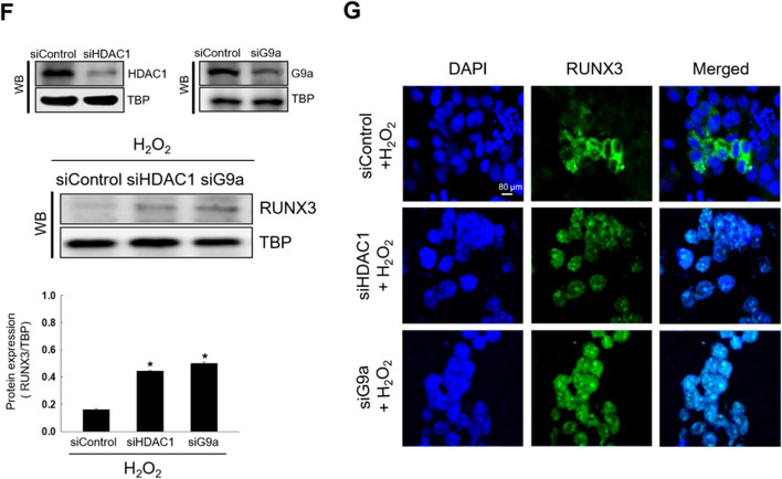
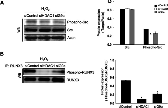
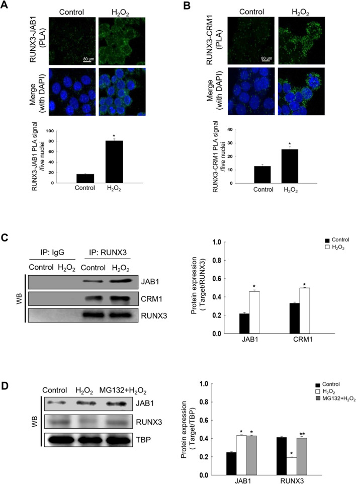
Runt domain transcription factor 3 (RUNX3) suppresses many different cancer types and is disabled by mutations, epigenetic repression, or cytoplasmic mislocalization. In this study, we investigated whether oxidative stress is associated with RUNX3 accumulation from the nucleus to the cytoplasm in terms of histone modification. Oxidative stress elevated histone deacetylase (HDAC) level and lowered that of histone acetyltransferase. In addition, oxidative stress decreased the expression of mixed lineage leukemia (MLL), a histone methyltransferase, but increased the expression of euchromatic histone-lysine N-methyltransferase 2 (EHMT2/G9a), which is also a histone methyltransferase. Moreover, oxidative stress-induced RUNX3 phosphorylation, Src activation, and Jun activation domain-binding protein 1 (JAB1) expression were inhibited by knockdown of HDAC and G9a, restoring the nuclear localization of RUNX3 under oxidative stress. Cytoplasmic RUNX3 localization was followed by oxidative stress-induced histone modification, activated Src along with RUNX3 phosphorylation, and induction of JAB1, resulting in RUNX3 inactivation.

Keywords: Cytoplasmic localization; Histone modification; Jun activation domain-binding protein 1; Oxidative stress; Runt domain transcription factor 3.


Conflict of interest statement

Declarations. Ethical Approval: Not applicable. Consent to Participate: Not applicable. Consent to Publication: Not applicable. Conflict of Interest: The authors declare no conflict of interest.

Figures

Fig. 1
Fig. 1
Oxidative stress-induced RUNX3 mislocalization by histone modification-related proteins. A Cell viability was assessed using MTT assay. *Significantly different from control cells (p < 0.05). Cells were pretreated with 1 mM NAC for 1 h, followed by treatment with H2O2 for 48 h. B ROS levels were determined using flow cytometry after H2DCFDA staining. *Significantly different from control cells (p < 0.05); **significantly different from H2O2-treated cells (p < 0.05). C Confocal microscopy images showed the RUNX3 protein in green fluorescence and nuclei in blue fluorescence. The merged image of H2O2-treated cells revealed the cytosolic localization of RUNX3. DNA was stained with DAPI. D, Western blot (WB) analysis was performed using antibodies against HDAC1, HAT1, EHMT2/G9a, MLL, and RUNX3. TBP was used as the loading control for nuclear protein fraction. D *Significantly different from 0 h of H2O2 treatment group (p < 0.05). *Significantly different from control cells (p < 0.05); **significantly different from H2O2-treated cells (p < 0.05). Cells were transfected with siHDAC1 RNA or siG9a RNA and treated with H2O2 for 48 h. F WB analysis for RUNX3 detection was performed. *Significantly different from H2O2 treated-siControl cells (p < 0.05). Confocal microscopy images showed the RUNX3 protein in green fluorescence, and nuclei in blue fluorescence. The merged image of siHDAC1 RNA- or siG9a RNA-transfected cells with H2O2 treatment indicated the nuclear localization of RUNX3
Fig. 1
Fig. 1
Oxidative stress-induced RUNX3 mislocalization by histone modification-related proteins. A Cell viability was assessed using MTT assay. *Significantly different from control cells (p < 0.05). Cells were pretreated with 1 mM NAC for 1 h, followed by treatment with H2O2 for 48 h. B ROS levels were determined using flow cytometry after H2DCFDA staining. *Significantly different from control cells (p < 0.05); **significantly different from H2O2-treated cells (p < 0.05). C Confocal microscopy images showed the RUNX3 protein in green fluorescence and nuclei in blue fluorescence. The merged image of H2O2-treated cells revealed the cytosolic localization of RUNX3. DNA was stained with DAPI. D, Western blot (WB) analysis was performed using antibodies against HDAC1, HAT1, EHMT2/G9a, MLL, and RUNX3. TBP was used as the loading control for nuclear protein fraction. D *Significantly different from 0 h of H2O2 treatment group (p < 0.05). *Significantly different from control cells (p < 0.05); **significantly different from H2O2-treated cells (p < 0.05). Cells were transfected with siHDAC1 RNA or siG9a RNA and treated with H2O2 for 48 h. F WB analysis for RUNX3 detection was performed. *Significantly different from H2O2 treated-siControl cells (p < 0.05). Confocal microscopy images showed the RUNX3 protein in green fluorescence, and nuclei in blue fluorescence. The merged image of siHDAC1 RNA- or siG9a RNA-transfected cells with H2O2 treatment indicated the nuclear localization of RUNX3
Fig. 2
Fig. 2
HDAC1 and G9a activate Src following oxidative stress. Cells were transfected with small interfering RNA HDAC1 or G9a and exposed to H2O2 for 48 h. A Levels of phospho-Src (the active form of Src) and Src were assessed using western blotting (WB). Actin was used as the loading control for total protein fraction. *Significantly different from H2O2 treated-siControl cells (p < 0.05). B Cellular lysates were subjected to immunoprecipitation (IP) using antibody against RUNX3, and subsequently examined by WB using antibody against phospho-tyrosine (phospho-RUNX3). *Significantly different from H2O2 treated-siControl cells (p < 0.05)
Fig. 3
Fig. 3
Oxidative stress induces JAB1 and CRM1 interaction via HDAC1 or G9a-induced Src activation. A Nuclear fraction was assessed using antibodies against JAB1, and CRM1. *Significantly different from control cells (p < 0.05), **significantly different from H2O2-treated cells (p < 0.05). B Cell lysates were immunoprecipitated with antibodies against JAB1 and CRM1 and subjected to WB. *Significantly different from control cells (p < 0.05). C The interaction between JAB1 and CRM1 was assessed using PLA; each green spot represented a single interaction. DNA was stained with DAPI. *Significantly different from control cells (p < 0.05). DWB was assessed for detection of nuclear JAB1 expression (D) in siHDAC1 RNA- or siG9a RNA-transfected cells with H2O2 treatment or (E) in Src-transfected cells with H2O2 treatment. D *Significantly different from H2O2 treated-siControl cells (p < 0.05). E *Significantly different from siControl cells (p < 0.05), **Significantly different from H2O2 treated-siControl cells (p < 0.05)
Fig. 4
Fig. 4
Oxidative stress induces RUNX3 mislocalization by inducing RUNX3, JAB1, and CRM1 interaction. A, B The interactions between (A) RUNX3 and JAB1 or (B) RUNX3 and CRM1 were assessed in H2O2-treated cells using PLA, and each green spot represented a single interaction. DNA was stained with DAPI. *Significantly different from control cells (p < 0.05). C The interactions of RUNX3 and JAB1 or RUNX3 and CRM1 were examined using IP with an antibody against RUNX3, followed by WB with antibodies against JAB1, CRM1, and RUNX3. Cells were pretreated with 20 µM MG132 for 1 h and then treated with H2O2 for 48 h. *Significantly different from control cells (p < 0.05). D Cell lysates were analyzed by WB with antibodies against JAB1 and RUNX3. Cells were transfected siJAB1 RNA or 10 nM leptomycin B and treated with H2O2 for 48 h. *Significantly different from control cells (p < 0.05); **significantly different from H2O2-treated cells (p < 0.05). E, F Confocal microscopy images showed RUNX3 protein in green fluorescence and nuclei in blue fluorescence. E The merged image of siJAB1 RNA-transfected cells with H2O2 treatment indicated the nuclear localization of RUNX3. F The merged image of leptomycin B-treated cells with H2O2 treatment indicates the nuclear localization of RUNX3
Fig. 4
Fig. 4
Oxidative stress induces RUNX3 mislocalization by inducing RUNX3, JAB1, and CRM1 interaction. A, B The interactions between (A) RUNX3 and JAB1 or (B) RUNX3 and CRM1 were assessed in H2O2-treated cells using PLA, and each green spot represented a single interaction. DNA was stained with DAPI. *Significantly different from control cells (p < 0.05). C The interactions of RUNX3 and JAB1 or RUNX3 and CRM1 were examined using IP with an antibody against RUNX3, followed by WB with antibodies against JAB1, CRM1, and RUNX3. Cells were pretreated with 20 µM MG132 for 1 h and then treated with H2O2 for 48 h. *Significantly different from control cells (p < 0.05). D Cell lysates were analyzed by WB with antibodies against JAB1 and RUNX3. Cells were transfected siJAB1 RNA or 10 nM leptomycin B and treated with H2O2 for 48 h. *Significantly different from control cells (p < 0.05); **significantly different from H2O2-treated cells (p < 0.05). E, F Confocal microscopy images showed RUNX3 protein in green fluorescence and nuclei in blue fluorescence. E The merged image of siJAB1 RNA-transfected cells with H2O2 treatment indicated the nuclear localization of RUNX3. F The merged image of leptomycin B-treated cells with H2O2 treatment indicates the nuclear localization of RUNX3

Similar articles

References

    1. Lotem, J., et al. (2017). Runx3 in immunity, inflammation and cancer. Advances in Experimental Medicine and Biology,962, 369–393. - PubMed
    1. Ito, Y., Bae, S. C., & Chuang, L. S. (2015). The RUNX family: Developmental regulators in cancer. Nature Reviews Cancer,15(2), 81–95. - PubMed
    1. Toska, A., Modi, N., & Chen, L. F. (2023). RUNX3 meets the ubiquitin-proteasome system in cancer. Cells,12(5), 717. - PMC - PubMed
    1. Tsang, Y. H., Lamb, A., & Chen, L. F. (2011). New insights into the inactivation of gastric tumor suppressor RUNX3: the role of H. pylori infection. Journal of Cellular Biochemistry,112(2), 381–386. - PMC - PubMed
    1. Zhou, X., et al. (2017). Mislocalization of Runt-related transcription factor 3 results in airway inflammation and airway hyper-responsiveness in a murine asthma model. Experimental and Therapeutic Medicine,14(3), 2695–2701. - PMC - PubMed

MeSH terms

LinkOut - more resources