. 2024 May 9;67(9):7443-7457.
doi: 10.1021/acs.jmedchem.4c00303. Epub 2024 Apr 29.

Identification of Potent and Selective Inhibitors of Acanthamoeba: Structural Insights into Sterol 14α-Demethylase as a Key Drug Target

Affiliations

Identification of Potent and Selective Inhibitors of Acanthamoeba: Structural Insights into Sterol 14α-Demethylase as a Key Drug Target

Tatiana Y Hargrove et al. J Med Chem. .

Abstract

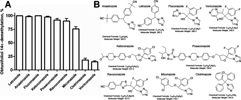
Acanthamoeba are free-living pathogenic protozoa that cause blinding keratitis, disseminated infection, and granulomatous amebic encephalitis, which is generally fatal. The development of efficient and safe drugs is a critical unmet need. Acanthamoeba sterol 14α-demethylase (CYP51) is an essential enzyme of the sterol biosynthetic pathway. Repurposing antifungal azoles for amoebic infections has been reported, but their inhibitory effects on Acanthamoeba CYP51 enzymatic activity have not been studied. Here, we report catalytic properties, inhibition, and structural characterization of CYP51 from Acanthamoeba castellanii. The enzyme displays a 100-fold substrate preference for obtusifoliol over lanosterol, supporting the plant-like cycloartenol-based pathway in the pathogen. The strongest inhibition was observed with voriconazole (1 h IC50 0.45 μM), VT1598 (0.25 μM), and VT1161 (0.20 μM). The crystal structures of A. castellanii CYP51 with bound VT1161 (2.24 Å) and without an inhibitor (1.95 Å), presented here, can be used in the development of azole-based scaffolds to achieve optimal amoebicidal effectiveness.


Conflict of interest statement

The authors declare no competing financial interest.

Figures

Figure 1
Figure 1
B-type CYP51 signature proline (marked red). (A) In the superimposed N. fowleri (cyan, 7RQT), T. brucei (plum, 3G1Q), and human (gray, 6UEZ) structures. (B) The proline-surrounding fragment of the aligned CYP51 sequences.
Figure 2
Figure 2
Optical absorbance of purified A. castellanii CYP51. The absolute spectrum of ferric protein and the difference spectrum of the ferrous CO-complex (inset). The P450 concentration is 10 μM. The absolute spectrum of the ferric sample was taken first, then the protein was reduced with sodium dithionite and the CO gas was bubbled through the cuvette after taking the baseline.
Figure 3
Figure 3
Catalytic activity of A. castellanii CYP51 toward obtusifoliol and lanosterol. (A) Time course of substrate conversion (37 °C, 0.5 μM P450, 2 μM CPR, 50 μM sterol). (B) Michaelis–Menten curves (calculated from the 1 min reactions with obtusifoliol and 20 min reactions with lanosterol). Rates (V0) are shown as nmol product formed per min per nmol P450. The experiments were performed in triplicate; the results are presented as mean ± SD. (C) Structural formulas.
Figure 4
Figure 4
Spectral responses to the binding of sterol substrates (A) obtusifoliol, (B) lanosterol, 2 μM P450, optical path 1 cm, and (C) a tetrazole-based inhibitor VT1161, 0.5 μM P450, optical path length 5 cm. Absolute absorption spectra before (black) and after the titrations (blue and red, respectively). Inset: The titration curves and the corresponding difference absorption spectra used in each analysis. Structural formulas are shown in Figure 6. The titration curves for the binding of other heterocyclic ligands are presented in Figure S2.
Figure 5
Figure 5
Clinical drugs. (A) Inhibition of A. castellanii CYP51 catalysis by commercial azole-based drugs in a 1 h reaction at the drug/enzyme molar ratio 3/1 (final concentrations 1.5/0.5 μM). The concentration of obtusifoliol was 50 μM. The experiments were performed in triplicate; the results are presented as mean ± SD. (B) Structural formulas.
Figure 6
Figure 6
Experimental CYP51 inhibitors. (A) Dose-dependent effects of imidazole-, pyridine-, and tetrazole-based CYP51 inhibitory scaffolds on the activity of the A. castellanii ortholog, 1 h reaction. Fluconazole and voriconazole were used as controls. P450 concentration was 0.5 μM; obtusifoliol concentration was 50 μM. The experiments were performed in triplicate; the results are presented as mean ± SD (B) structural formulas.
Figure 7
Figure 7
Binding mode of n-dodecyl-β-d-maltoside. (7UWP). (A) Inside the active site. The heme-bound water molecule is shown as a black sphere. The detergent (tan carbons) is in ball and stick representation, and the residues within 4.5 Å from its fatty acid tail are shown as sticks. The corresponding secondary structural elements are pink, and the rest of the ribbon is semitransparent. The FG arm (195–256) is plum. Inset: the detergent structural formula. (B) Outside the protein globule. Surface representation. The detergent-formed channel between helices F, I, and the β4 hairpin is seen through the semitransparent surface. The proton relay salt bridge (D213-H297) is on the right.
Figure 8
Figure 8
Heme surrounding in A. castellanii CYP51 (7UWP). Six protein residues forming interactions with the heme are labeled. Hydrogen bonds are shown as dashed green lines and iron coordination bonds are presented as orange springs. The secondary structural elements are seen as semitransparent ribbons.
Figure 9
Figure 9
Size-exclusion chromatography on Superdex 200 increase 10/300 GL. (A) Column calibration in PBS and 0.5 mL/min flow rate. (B) The HPLC profile of the A. castellanii CYP51 sample. (C) Absolute absorption spectra of peaks 1 (dimer) and 2 (monomer).
Figure 10
Figure 10
VT1161 binding mode (8EKT). (A) Inside the active site of A. castellanii CYP51. The inhibitor (magenta carbons), the heme, and amino acid residues within 4.5 Å (blue carbons) are shown as sticks, and the corresponding secondary structural elements are seen as semitransparent ribbons. Y114, F293, and M471 (pink carbons) are from the superimposed inhibitor-free structure. (B) Superimposition of 8EKT with VT1161-bound T. cruzi (brown, 5AJR) and C. albicans (yellow, 5TZ1) CYP51s. F48/Y64 are from helix A′ (F60 in A. castellanii); Y116 in complex with VT1161 in T. cruzi CYP51 loses its H-bond with the heme propionate (Y127 in A. castellanii). H377 in C. albicans forms a H-bond with the trifluoroethoxy oxygen of the inhibitor (L364 in A. castellanii). Hydrogen bonds are shown as dashed green lines and iron coordination bonds are presented as orange springs. The secondary structural elements are seen as semitransparent ribbons.
Figure 11
Figure 11
Voriconazole docked in the 7UWP structure of A. castellanii CYP51. The inhibitor (dark red carbons), the heme, Y114, and T298 (light green carbons) are shown as sticks and possible H-bonds are depicted as dashed green lines.
Figure 12
Figure 12
Substrate-gating residue in the B′ helix. A. castellanii CYP51 (8EKT, tan) is superimposed with the structures of T. brucei (plum, 3G1Q), N. fowleri (cyan, 7RTQ), and lanosterol (green)-bound human (semitransparent gray, 6UEZ) orthologs. Membrane-oriented side of the molecule. Inset: Enlarged view of A. castellanii F116 and lanosterol in sphere representation; the C4 atom α and β methyl groups (C28 and C29 carbons) are marked. The clash between the 4β methyl group of lanosterol and F116 indicates that in A. castellanii CYP51, a structural rearrangement in the B′ helix is required for this substrate to reach the catalytic site.

Similar articles

Cited by

References

    1. Guimaraes A. J.; Gomes K. X.; Cortines J. R.; Peralta J. M.; Peralta R. H. Acanthamoeba spp. as a Universal Host for Pathogenic Microorganisms: One Bridge from Environment to Host Virulence. Microbiol. Res. 2016, 193, 30–38. 10.1016/j.micres.2016.08.001. - DOI - PubMed
    1. Ashbolt N. J. Environmental (Saprozoic) Pathogens of Engineered Water Systems: Understanding Their Ecology for Risk Assessment and Management. Pathogens 2015, 4 (2), 390–405. 10.3390/pathogens4020390. - DOI - PMC - PubMed
    1. Lorenzo-Morales J.; Khan N. A.; Walochnik J. An Update on Acanthamoeba keratitis: Diagnosis, Pathogenesis and Treatment. Parasite 2015, 22, 1010.1051/parasite/2015010. - DOI - PMC - PubMed
    1. Aichelburg A. C.; Walochnik J.; Assadian O.; Prosch H.; Steuer A.; Perneczky G.; Visvesvara G. S.; Aspöck H.; Vetter N. Successful Treatment of Disseminated Acanthamoeba sp. Infection with Miltefosine. Emerging Infect. Dis. 2008, 14 (11), 1743–1746. 10.3201/eid1411.070854. - DOI - PMC - PubMed
    1. Baig A. M. Granulomatous Amoebic Encephalitis: Ghost Response of an Immunocompromised Host?. J. Med. Microbiol. 2014, 63 (Pt 12), 1763–1766. 10.1099/jmm.0.081315-0. - DOI - PubMed

Publication types

MeSH terms

Substances