. 2024 May 8;146(18):12702-12711.
doi: 10.1021/jacs.4c02262. Epub 2024 Apr 29.

A Targetable N-Terminal Motif Orchestrates α-Synuclein Oligomer-to-Fibril Conversion

Affiliations

A Targetable N-Terminal Motif Orchestrates α-Synuclein Oligomer-to-Fibril Conversion

Jaime Santos et al. J Am Chem Soc. .

Abstract

Oligomeric species populated during α-synuclein aggregation are considered key drivers of neurodegeneration in Parkinson's disease. However, the development of oligomer-targeting therapeutics is constrained by our limited knowledge of their structure and the molecular determinants driving their conversion to fibrils. Phenol-soluble modulin α3 (PSMα3) is a nanomolar peptide binder of α-synuclein oligomers that inhibits aggregation by blocking oligomer-to-fibril conversion. Here, we investigate the binding of PSMα3 to α-synuclein oligomers to discover the mechanistic basis of this protective activity. We find that PSMα3 selectively targets an α-synuclein N-terminal motif (residues 36-61) that populates a distinct conformation in the mono- and oligomeric states. This α-synuclein region plays a pivotal role in oligomer-to-fibril conversion as its absence renders the central NAC domain insufficient to prompt this structural transition. The hereditary mutation G51D, associated with early onset Parkinson's disease, causes a conformational fluctuation in this region, leading to delayed oligomer-to-fibril conversion and an accumulation of oligomers that are resistant to remodeling by molecular chaperones. Overall, our findings unveil a new targetable region in α-synuclein oligomers, advance our comprehension of oligomer-to-amyloid fibril conversion, and reveal a new facet of α-synuclein pathogenic mutations.


Conflict of interest statement

The authors declare the following competing financial interest(s): SV, IP and JS have submitted a patent protecting the use of PSM3 for therapy and diagnosis. Request number: EP20382658. Priority date: 22-07-2020.

Figures

Figure 1
Figure 1
PSMα3 binding to αS oligomers. (a) Schematic representation of PSMα3 binding and activities. (b) Cross-linking map representing PSMα3 contacts with αS oligomers. (c) Wood’s plots showing the difference in deuterium uptake (ΔDU) when comparing αS oligomers in the complex with PSMα3 and free αS oligomers by HDX-MS at the 60 s exposure time point. Peptides colored blue are protected from exchange in the presence of PSMα3 (see the Experimental Section), suggesting that they are less solvent-exposed and/or participate in more inter/intraprotein hydrogen bonding in the presence of PSMα3. (d) 2D 13C–13C PDSD correlation spectra (mixing time of 50 ms) of oligomers (black) and oligomers + PSMα3 (green). (e) 3D reconstruction of αS oligomers in the absence of PSMα3 (18.5 Å resolution). (f) 3D reconstruction of αS oligomers in the complex with PSMα3 (19 Å resolution).
Figure 2
Figure 2
Dynamics of the N-terminal region of αS in the oligomer. (a) Wood’s plots showing the difference in deuterium uptake (ΔDU) between αS monomers and oligomers by HDX-MS at the 60 s exposure time point to deuterium. Peptides colored blue are significantly protected from exchange in αS oligomers compared with monomeric αS. (b) Two views of the SAXS-based 3D reconstruction of αS oligomers. The compact core (blue) is surrounded by an outer disordered shell (green). The cryoEM density map is shown inside the oligomer core (gray).
Figure 3
Figure 3
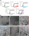
Contribution of PSMα3 binding site to oligomer-to-fibril conversion. (a) Kinetics of amyloid formation of the WT, ΔP1, ΔP2, ΔΔ, Y39A, and S42A variants monitored using Th-T fluorescence. (b) Representative nsEM images of the oligomeric fraction of the WT (top left), ΔP1 (top middle), ΔP2 (top right), ΔΔ (bottom left), Y39A (bottom middle), and S42A (bottom right) isolated at the end point (WT, ΔP1, ΔP2, ΔΔ, and Y39A) or after 28 h of assembly (S42A).
Figure 4
Figure 4
Effect of the familial G51D mutation on αS amyloid and oligomer formation and disaggregation by molecular chaperones. (a) Wood’s plot showing the relative solvent exposure/hydrogen bonding of G51D αS oligomers compared with that of WT αS oligomers by HDX-MS at the 60 s time point of exposure to deuterium. Deprotection from deuterium uptake occurs in the N-terminal region, as indicated by the peptide region colored red. (b) Assembly kinetics of G51D into amyloid fibrils monitored using ThT fluorescence. (c) Representative nsEM micrographs of the G51D oligomeric fraction after 28 h of assembly. (d,e) Sucrose-gradient fractionation of WT (d) and G51D (e) oligomers in the absence (left panels) or upon 2.5 h of incubation at 30 °C with the human disaggregase at αS/Hsc70 1:1.5 molar ratios (central panels). The distribution across the gradient was followed by Western blot analysis using an anti-αS antibody. The relative intensity of the immunoreactive bands in the Western blots was quantified for nontreated (dotted line) and treated (solid line) oligomers to illustrate their differential distribution (right panels).
Figure 5
Figure 5
Schematic representation of the αS aggregation landscape.

References

    1. Spillantini M. G.; Schmidt M. L.; Lee V. M.; Trojanowski J. Q.; Jakes R.; Goedert M. α-Synuclein in Lewy bodies. Nature 1997, 388 (6645), 839–840. 10.1038/42166. - DOI - PubMed
    1. Polymeropoulos M. H.; Lavedan C.; Leroy E.; Ide S. E.; Dehejia A.; Dutra A.; Pike B.; Root H.; Rubenstein J.; Boyer R.; Stenroos E. S.; Chandrasekharappa S.; Athanassiadou A.; Papapetropoulos T.; Johnson W. G.; Lazzarini A. M.; Duvoisin R. C.; Di Iorio G.; Golbe L. I.; Nussbaum R. L. Mutation in the α-Synuclein Gene Identified in Families with Parkinson’s Disease. Science 1997, 276 (5321), 2045–2047. 10.1126/science.276.5321.2045. - DOI - PubMed
    1. Goedert M.; Jakes R.; Spillantini M. G. The Synucleinopathies: Twenty Years On. J. Park. Dis. 2017, 7 (s1), S51–S69. 10.3233/JPD-179005. - DOI - PMC - PubMed
    1. Cremades N.; Cohen S. I. A.; Deas E.; Abramov A. Y.; Chen A. Y.; Orte A.; Sandal M.; Clarke R. W.; Dunne P.; Aprile F. A.; Bertoncini C. W.; Wood N. W.; Knowles T. P. J.; Dobson C. M.; Klenerman D. Direct Observation of the Interconversion of Normal and Toxic Forms of α-Synuclein. Cell 2012, 149 (5), 1048–1059. 10.1016/j.cell.2012.03.037. - DOI - PMC - PubMed
    1. Zurlo E.; Kumar P.; Meisl G.; Dear A. J.; Mondal D.; Claessens M. M. A. E.; Knowles T. P. J.; Huber M. In Situ Kinetic Measurements of α-Synuclein Aggregation Reveal Large Population of Short-Lived Oligomers. PLoS One 2021, 16 (1), e024554810.1371/journal.pone.0245548. - DOI - PMC - PubMed

Publication types

Substances