. 2024 May 7;121(19):e2313590121.
doi: 10.1073/pnas.2313590121. Epub 2024 Apr 29.

Neuronal innervation regulates the secretion of neurotrophic myokines and exosomes from skeletal muscle

Affiliations

Neuronal innervation regulates the secretion of neurotrophic myokines and exosomes from skeletal muscle

Kai-Yu Huang et al. Proc Natl Acad Sci U S A. .

Abstract

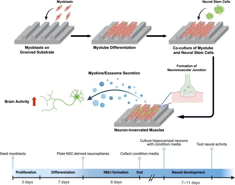
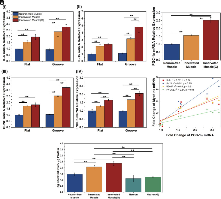
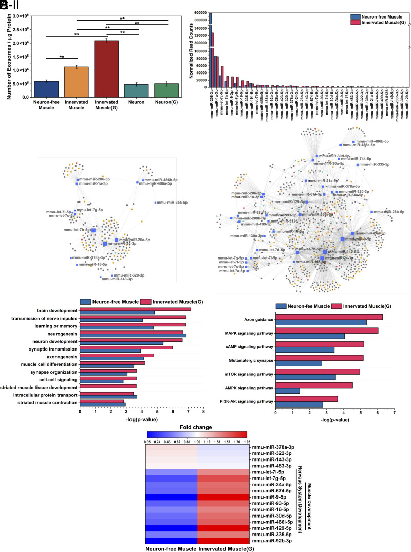
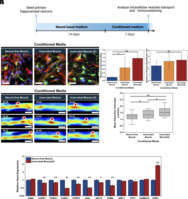
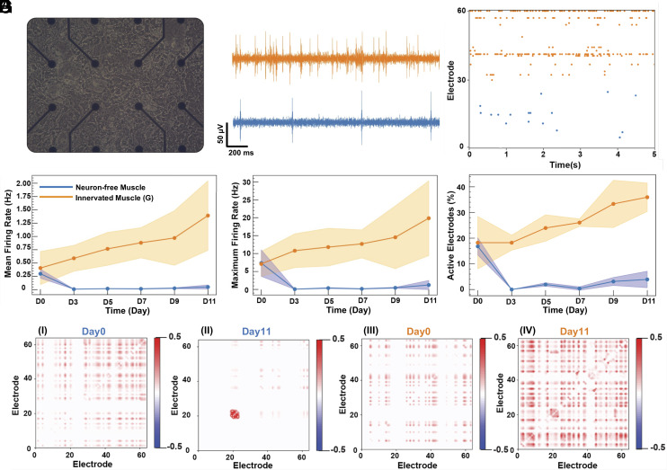
Myokines and exosomes, originating from skeletal muscle, are shown to play a significant role in maintaining brain homeostasis. While exercise has been reported to promote muscle secretion, little is known about the effects of neuronal innervation and activity on the yield and molecular composition of biologically active molecules from muscle. As neuromuscular diseases and disabilities associated with denervation impact muscle metabolism, we hypothesize that neuronal innervation and firing may play a pivotal role in regulating secretion activities of skeletal muscles. We examined this hypothesis using an engineered neuromuscular tissue model consisting of skeletal muscles innervated by motor neurons. The innervated muscles displayed elevated expression of mRNAs encoding neurotrophic myokines, such as interleukin-6, brain-derived neurotrophic factor, and FDNC5, as well as the mRNA of peroxisome-proliferator-activated receptor γ coactivator 1α, a key regulator of muscle metabolism. Upon glutamate stimulation, the innervated muscles secreted higher levels of irisin and exosomes containing more diverse neurotrophic microRNAs than neuron-free muscles. Consequently, biological factors secreted by innervated muscles enhanced branching, axonal transport, and, ultimately, spontaneous network activities of primary hippocampal neurons in vitro. Overall, these results reveal the importance of neuronal innervation in modulating muscle-derived factors that promote neuronal function and suggest that the engineered neuromuscular tissue model holds significant promise as a platform for producing neurotrophic molecules.

Keywords: exosome; innervation; myokine; neuromuscular junction; skeletal muscle.


Conflict of interest statement

Competing interests statement:The authors declare no competing interest.

Figures

Fig. 1.
Fig. 1.
Schematic of creating a neuron-innervated muscle model using a grooved pattern substrate and subsequent analysis of muscle secretory activity. Created with BioRender.com.
Fig. 2.
Fig. 2.
Effect of substrate topology on recreation of neuron-innervated muscle. (A) Immunofluorescence images of myotubes (MHC, red) and nucleus (DAPI, blue) of primary myoblasts differentiated on flat and microgrooved (Groove) substrates. (B) MHC-positive area divided by total cell number and (C) fusion index of C2C12-derived or primary myoblast-derived myotubes. (n = 5, *P < 0.05). (D) Immunofluorescence images of the neuron-innervated muscle made with primary myoblasts. [myotubes (MHC, red), neurons (TUBB3, green), AChRs (α-BTX, yellow), and nuclei (DAPI, blue)] (E) Innervation of neuron on primary myoblast-derived myotube on a grooved substrate. The white arrows indicate the innervation spot of the axonal terminal toward AChR clusters. (F) Relative AChR expression area on myotubes. (G) Relative colocalization area of axon and AChR on innervated muscles. (n ≥ 3, *P < 0.05, **P < 0.01). (H) Scanning electron microscopy (SEM) images of the innervated muscle.
Fig. 3.
Fig. 3.
Analysis of muscle contraction of Neuron-free Muscle, Innervated Muscle, and Innervated Muscle(G), created on a flat and a microgrooved (Groove) substrate. (A) Visualization of the muscle contraction on the flat and the grooved substrate. Size of the view: 299.5 μm × 224.6 μm. The z value represented the number of contractions in each block per second. (B) Percentage of contracting myotubes. (C) Relative contraction displacement of myotubes. The “y” value was normalized to the value of Neuron-free Muscle on the flat substrate. (D) Direction of muscle contraction 90 and 270 degrees were aligned with the grooved pattern direction. (E) Average contraction frequency of the muscles (n= 3, *P < 0.05, **P < 0.01).
Fig. 4.
Fig. 4.
Effect of neuronal innervation on myokine-encoding mRNA expression and irisin secretion of skeletal muscles. Primary myoblasts were used in this analysis. (A) IL-6 (I), IL-15 (II), BDNF (III), and FNDC5 (IV)-encoding mRNA levels of Neuron-free Muscle, Innervated Muscle, and Innervated Muscle(G) on the flat and the microgrooved substrates after 8 d of coculture. mRNA levels were quantified with a fold change to the expression of Neuron-free Muscle formed on the flat substrate. (B) PGC-1α-encoding mRNA expression of muscles on the grooved substrate. (C) Correlation of IL-6, IL-15, BDNF, and FNDC5-encoding mRNA expressions to PGC-1α-encoding mRNA expression of the muscles on the grooved substrates. (r2: R-squared value, p: correlation coefficient) (D) Extracellular irisin concentration normalized to the total intracellular protein level of muscles or NSC-derived neurons (Neuron) and NSC-derived neurons incubated with 500 µM glutamate [Neuron(G)] on the microgrooved substrate. The protein concentration was measured after 8 d of coculture (n = 3, *P < 0.05, **P < 0.01).
Fig. 5.
Fig. 5.
Analysis of exosomes released from neuron-free muscle and innervated muscle. (A) Number of exosomes in the conditioned media normalized to intracellular proteins after 24-h incubation (n = 3, *P < 0.05, **P < 0.01). (B) Normalized read counts of the top 95% abundant exosomal miRNAs. (C) miRNA-target gene network of the top 95% miRNAs of (C, I) Neuron-free Muscle and (C, II) Innervated Muscle(G). Genes targeted by at least two miRNAs were selected for the network analysis. Genes related to nervous system development were marked with a yellow circle, and muscle development-related genes were marked with red circles. Genes regulating both nerve and muscle development were marked with green circles. (D) Enriched nervous system and muscle development-related biological processes and (E) Enriched KEGG pathways regulated by the top 95% miRNA in the exosomes secreted by Neuron-free Muscle or Innervated Muscle(G). (F) Heat map showing the fold change of top 95% exosomal miRNA expression between the Neuron-free Muscle and Innervated Muscle(G) (P-value < 0.05, n = 3). The color key indicates the fold change of miRNA expression; red indicates upregulation, and blue indicates downregulation.
Fig. 6.
Fig. 6.
Effects of muscle-derived conditioned media on neural development and intracellular axon transport. (A) Timeline of testing effects of muscle-derived conditioned media on hippocampal neurogenesis. Three different conditioned media were harvested from Neuron-free Muscle, Innervated Muscle, and Innervated Muscle(G) cultured on the microgrooved substrate. (B) Immunofluorescence images of hippocampal neurons cultured with conditioned media for 7 d. [neurons (MAP2, green), astrocytes (GFAP, red), and nuclei (DAPI, blue)] (C) Neuron-to-astrocyte ratio and (D) number of primary dendrites per cell altered with three different conditioned media (n > 5 per group, *P < 0.05, **P < 0.01). (E) Representative SLIM-based images of vesicular transport in neurites incubated with conditioned media (minutes:seconds). Red arrows indicated the tracked vesicles. (F) Mean advection velocities of vesicles transported in neurites quantified from SLIM images (n > 40 neurites, *P < 0.05, **P < 0.01). (G) Comparative gene expression analysis in hippocampal neurons after 7 d culture with conditioned media from Innervated Muscle(G) versus Neuron-free Muscle (n = 3, *P < 0.05, **P < 0.01).
Fig. 7.
Fig. 7.
Effects of muscle-derived conditioned media on neural electrophysiology. (A) Bright-field image of hippocampal neurons on MEA after 14 d of culture in the neurobasal medium. (B) Firing signals from one electrode of MEA. These signals were captured after incubating the neurons with conditioned media derived from Innervated Muscle(G) (orange) and Neuron-free Muscle (blue) for 11 d. (C) The raster plots of recording from each MEA with neurons incubated with the Innervated Muscle(G) medium (orange, electrode number 31 to 60) and Neuron-free Muscle medium (blue, electrode number 0 to 30) for 11 d. (D) Mean firing rate, (E) maximum firing rate, and (F) percentage of active electrodes from the neurons cultured with Neuron-free Muscle medium (blue) and Innervated Muscle(G) medium (orange) (n = 3). The data were analyzed from day 0 to day 11 after starting the culture of neurons with conditioned media. (G) Correlation between electrodes with neurons incubated in Neuron-free Muscle medium (G, I at day 0 and G, II at day 11) and Innervated Muscle(G) medium (G, III at day 0 and G, IV at day 11).

References

    1. Severinsen M. C. K., Pedersen B. K., Muscle-organ crosstalk: The Emerging roles of myokines. Endocrine Rev. 41, 594–609 (2020). - PMC - PubMed
    1. Pedersen B. K., Febbraio M. A., Muscle as an endocrine organ: Focus on muscle-derived interleukin-6. Physiol. Rev. 88, 1379–1406 (2008). - PubMed
    1. León-Ariza H. H., Mendoza-Navarrete M. P., Maldonado-Arango M. I., Botero-Rosas D. A., A systematic review of “myokines and metabolic regulation”. Apunts. Medicina de l’Esport 53, 155–162 (2018).
    1. Kim S., et al. , Roles of myokines in exercise-induced improvement of neuropsychiatric function. Pflügers Arch. 471, 491–505 (2019). - PubMed
    1. Wrann C. D., et al. , Exercise induces hippocampal BDNF through a PGC-1α/FNDC5 pathway. Cell Metab. 18, 649–659 (2013). - PMC - PubMed

Publication types

MeSH terms

LinkOut - more resources