. 2024 Apr 29;10(1):201.
doi: 10.1038/s41420-024-01967-8.

Emetine induces oxidative stress, cell differentiation and NF-κB inhibition, suppressing AML stem/progenitor cells

Affiliations

Emetine induces oxidative stress, cell differentiation and NF-κB inhibition, suppressing AML stem/progenitor cells

Suellen L R Silva et al. Cell Death Discov. .

Abstract

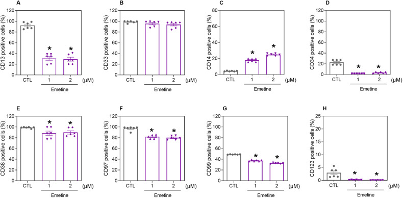
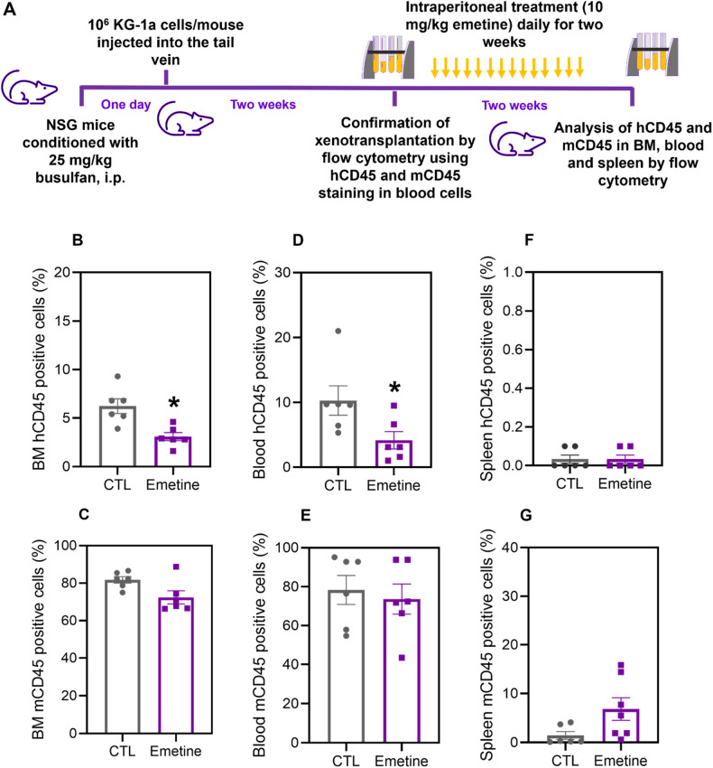
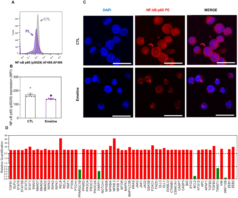
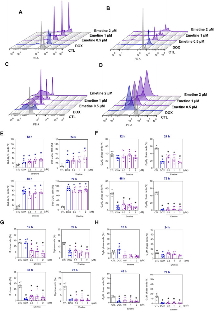
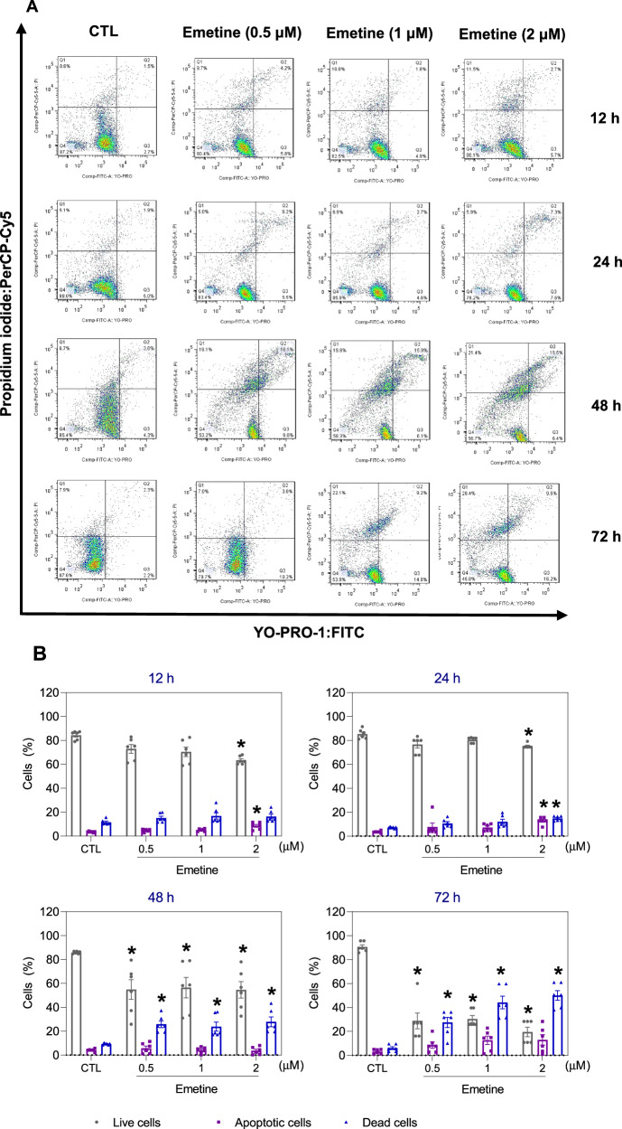
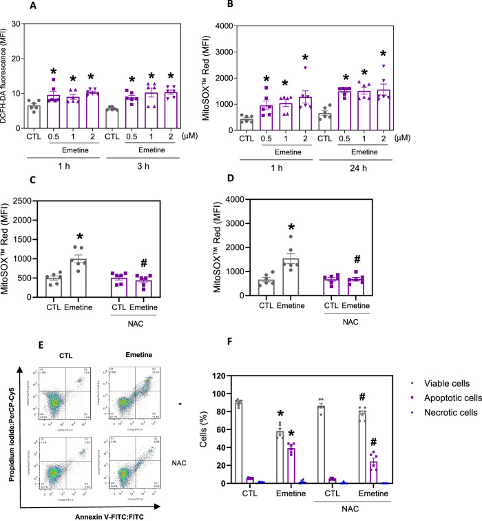
Acute myeloid leukemia (AML) is a fatal malignancy of the blood and bone marrow. Leukemic stem cells (LSCs) are a rare subset of leukemic cells that promote the development and progression of AML, and eradication of LSCs is critical for effective control of this disease. Emetine is an FDA-approved antiparasitic drug with antitumor properties; however, little is known about its potential against LSCs. Herein, we explored the antileukemic potential of emetine, focusing on its effects on AML stem/progenitor cells. Emetine exhibited potent cytotoxic activity both in hematologic and solid cancer cells and induced AML cell differentiation. Emetine also inhibited AML stem/progenitor cells, as evidenced by decreased expression of CD34, CD97, CD99, and CD123 in KG-1a cells, indicating anti-AML stem/progenitor cell activities. The administration of emetine at a dosage of 10 mg/kg for two weeks showed no significant toxicity and significantly reduced xenograft leukemic growth in vivo. NF-κB activation was reduced in emetine-treated KG-1a cells, as shown by reduced phospho-NF-κB p65 (S529) and nuclear NF-κB p65. DNA fragmentation, YO-PRO-1 staining, mitochondrial depolarization and increased levels of active caspase-3 and cleaved PARP (Asp214) were detected in emetine-treated KG-1a cells. Moreover, treatment with the pancaspase inhibitor Z-VAD(OMe)-FMK partially prevented the apoptotic cell death induced by emetine. Emetine treatment also increased cellular and mitochondrial reactive oxygen species, and emetine-induced apoptosis in KG-1a cells was partially prevented by the antioxidant N-acetylcysteine, indicating that emetine induces apoptosis, at least in part, by inducing oxidative stress. Overall, these studies indicate that emetine is a novel potential anti-AML agent with promising activity against stem/progenitor cells, encouraging the development of further studies aimed at its clinical application.


Conflict of interest statement

The authors declare no competing interests.

Figures

Fig. 1
Fig. 1. Emetine affects the viability of hematological and solid cancer cells.
A Chemical structure of emetine. B IC50 values of the cytotoxicity of emetine against hematological (red bars) and solid cancers (blue bars), as well as against noncancerous cells (green bars). C Heatmap of selectivity indixes (SI) calculated for emetine using the following formula: SI = IC50 [noncancer cells]/IC50 [cancer cells].
Fig. 2
Fig. 2. Emetine causes cell differentiation and suppresses AML stem/progenitor cells.
Immunophenotypic analysis of the myeloid lineage markers CD13 (A) and CD33 (B), the AML cell differentiation marker CD14 (C), and the AML stem/progenitor markers CD34 (D), CD38 (E), CD97 (F), CD99 (G), and CD123 (H) was performed in emetine-treated KG-1a cells after 48 h of incubation. The vehicle (0.2% DMSO) was used as a negative control (CTL). The data are shown as the mean ± S.E.M. of three independent experiments carried out in duplicate. *p < 0.05 compared to CTL by one-way ANOVA followed by Dunnett’s multiple comparisons test.
Fig. 3
Fig. 3. Effect of emetine on the growth of xenografts derived from KG-1a cells.
A A xenograft model was established in NSG mice. Two weeks after the inoculation of KG-1a cells, the mice were randomly divided into the emetine (10 mg/kg) group and the control group (5% DMSO). hCD45-positive cells were quantified by flow cytometry from (B) bone marrow, (D) peripheral blood and (F) spleen. mCD45-positive cells were quantified by flow cytometry from (C) bone marrow, (E) peripheral blood, and (G) spleen. The data are shown as the mean ± S.E.M. of 6 animals. *p < 0.05 compared with CTL by Student’s t test.
Fig. 4
Fig. 4. Emetine interferes with NF-κB signaling in KG-1a cells.
A, B Effect of emetine on the levels of NF-κB p65 (pS529) after 24 h of treatment in KG-1a cells. The cells were treated with 2 μM emetine. The vehicle (0.2% DMSO) was used as a negative control (CTL). The data are shown as the mean ± S.E.M. of three independent experiments carried out in duplicate. *p < 0.05 compared with CTL by Student’s t test. MFI = mean fluorescence intensity. C Representative immunofluorescence images of NF-κB p65 in KG-1a cells after 24 h of incubation with 2 μM emetine. Scale bar = 25 μm. D Up- and downregulated genes in KG-1a cells after 12 h of treatment with 2 µM emetine. Genes that displayed RQ ≥ 2 (red bars) were upregulated, and RQs ≤ 0.5 (green bars) were downregulated.
Fig. 5
Fig. 5. Cell cycle progression in KG-1a cells after incubation with emetine.
Representative histograms after (A) 12, (B) 24, (C) 48, and (D) 72 h of treatment. Percentages of cells in (E) sub-G0/G1, (F) G0/G1, (G) S, and (H) G2/M after different incubation periods with emetine. Vehicle (0.2% DMSO) was used as a negative control (CTL), and doxorubicin (DOX, 1 µM) was used as a positive control. The data are shown as the mean ± S.E.M. of three independent experiments carried out in duplicate. *p < 0.05 compared with CTL by one-way ANOVA followed by Dunnett’s multiple comparisons test.
Fig. 6
Fig. 6. Apoptotic cell death induced by emetine in KG-1a cells.
A Representative flow cytometry dot plots. B Apoptosis quantification in KG-1a cells after 12, 24, 48, and 72 h of treatment with emetine. Quantification of live (YO-PRO-1- and PI-double negative), apoptotic (YO-PRO-1-positive) and dead (YO-PRO-1- and PI-double positive) KG-1a cells. Vehicle (0.2% DMSO) was used as a negative control (CTL). The data are shown as the mean ± S.E.M. of three independent experiments carried out in duplicate. *p < 0.05 compared with CTL by one-way ANOVA followed by Dunnett’s multiple comparisons test.
Fig. 7
Fig. 7. Emetine causes caspase-mediated apoptosis in KG-1a cells.
A Effect of emetine on the levels of active caspase 3 and (B) cleaved PARP (Asp214) after 24 h of treatment in KG-1a cells. C Effect of emetine on mitochondrial activity in KG-1a cells. D, E Effect of the pancaspase inhibitor Z-VAD(OMe)-FMK on the apoptosis induced by emetine in KG-1a cells. The cells were pretreated for 2 h with 50 μM Z-VAD(OMe)-FMK and then incubated with 2 μM emetine for 48 h. F, G Survival curves of WT SV40 MEFs and BAD KO SV40 MEFs upon treatment with 5-fluorouracil (5-FU, a positive control) and emetine. The curves were obtained from at least three independent experiments carried out in duplicate using the Alamar blue assay after 72 h of incubation. H, I Induction of cell death in WT SV40 MEFs and BAD KO SV40 MEFs after 48 h of incubation with 2 μM emetine. Vehicle (0.2% DMSO) was used as a negative control (CTL), and doxorubicin (DOX, 1 µM) was used as a positive control. The data are shown as the mean ± S.E.M. of three independent experiments carried out in duplicate. *p < 0.05 compared with CTL by Student’s t test. #p < 0.05 compared with the respective treatment without inhibitor by Student’s t test. MFI mean fluorescence intensity.
Fig. 8
Fig. 8. Induced oxidative stress in KG-1a cells by emetine.
A Cellular ROS in KG-1a cells after 1 and 3 h of treatment with emetine. B Mitochondrial ROS in KG-1a cells after 1 and 24 h of treatment with emetine. Mitochondrial ROS in KG-1a cells after 1 (C) and 24 (D) h of treatment with 2 μM emetine pretreated with the antioxidant NAC. E, F Effect of the antioxidant NAC on the apoptosis induced by emetine in KG-1a cells. The cells were pretreated for 2 h with 5 mM NAC and then incubated with 2 μM emetine for 48 h. Vehicle (0.2% DMSO) was used as a negative control (CTL). The data are shown as the mean ± S.E.M. of three independent experiments carried out in duplicate. *p < 0.05 compared with CTL by Student’s t test or one-way ANOVA followed by Dunnett’s multiple comparisons test. #p < 0.05 compared with the respective treatment without inhibitor by Student’s t test. MFI mean fluorescence intensity.

Similar articles

Cited by

References

    1. Kantarjian H, Kadia T, DiNardo C, Daver N, Borthakur G, Jabbour E, et al. Acute myeloid leukemia: current progress and future directions. Blood Cancer J. 2021;11:41. doi: 10.1038/s41408-021-00425-3. - DOI - PMC - PubMed
    1. Döhner H, Wei AH, Appelbaum FR, Craddock C, DiNardo CD, Dombret H, et al. Diagnosis and management of AML in adults: 2022 recommendations from an international expert panel on behalf of the ELN. Blood. 2022;140:1345–77. doi: 10.1182/blood.2022016867. - DOI - PubMed
    1. DiNardo CD, Erba HP, Freeman SD, Wei AH. Acute myeloid leukaemia. Lancet. 2023;401:2073–86. doi: 10.1016/S0140-6736(23)00108-3. - DOI - PubMed
    1. American Cancer Society. Cancer facts & figures 2023. Atlanta: American Cancer Society; 2023.
    1. Bhansali RS, Pratz KW, Lai C. Recent advances in targeted therapies in acute myeloid leukemia. J Hematol Oncol. 2023;16:29. doi: 10.1186/s13045-023-01424-6. - DOI - PMC - PubMed