. 2024 Apr 29;15(1):3642.
doi: 10.1038/s41467-024-48029-2.

A secondary mechanism of action for triazole antifungals in Aspergillus fumigatus mediated by hmg1

Affiliations

A secondary mechanism of action for triazole antifungals in Aspergillus fumigatus mediated by hmg1

Jeffrey M Rybak et al. Nat Commun. .

Abstract

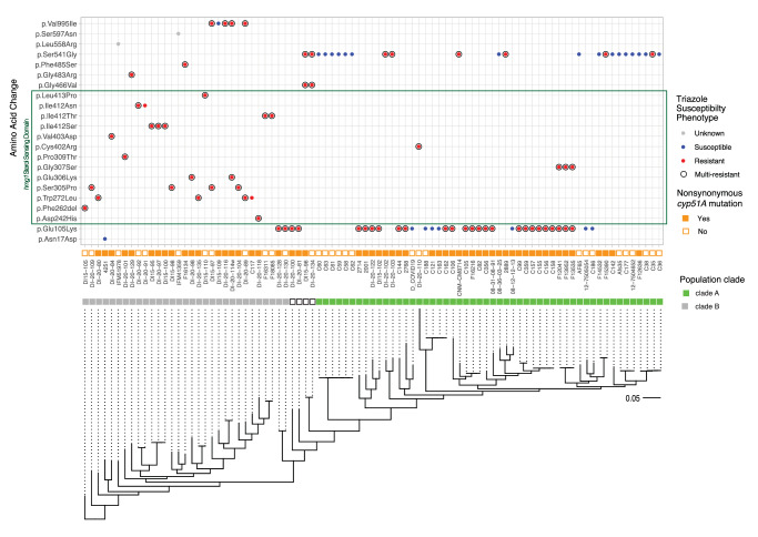
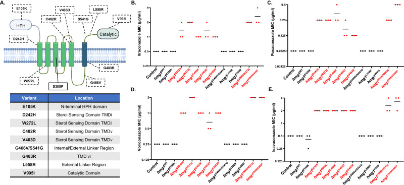
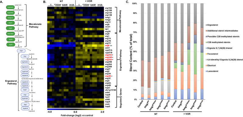
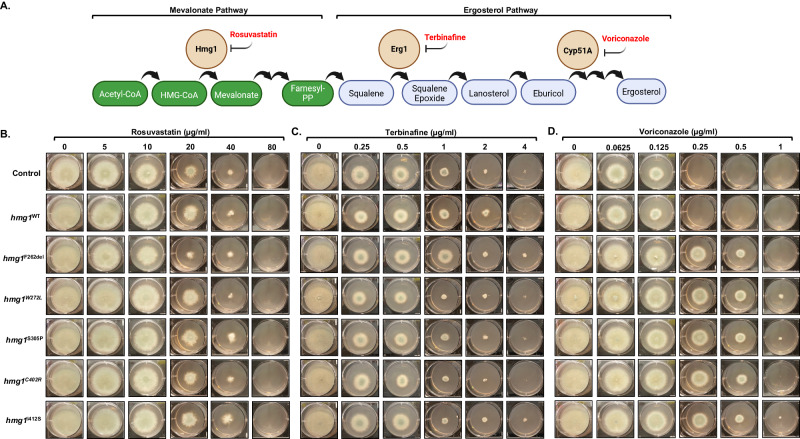
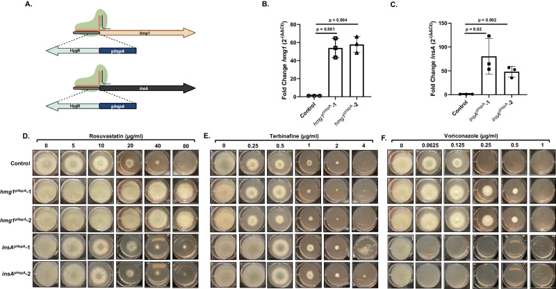
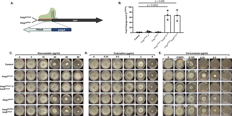
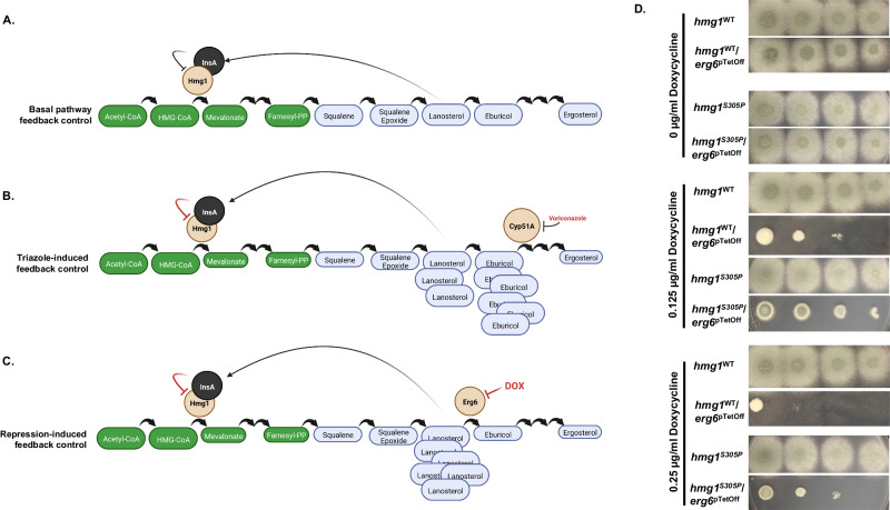
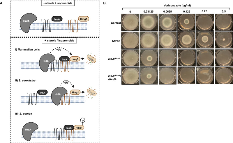
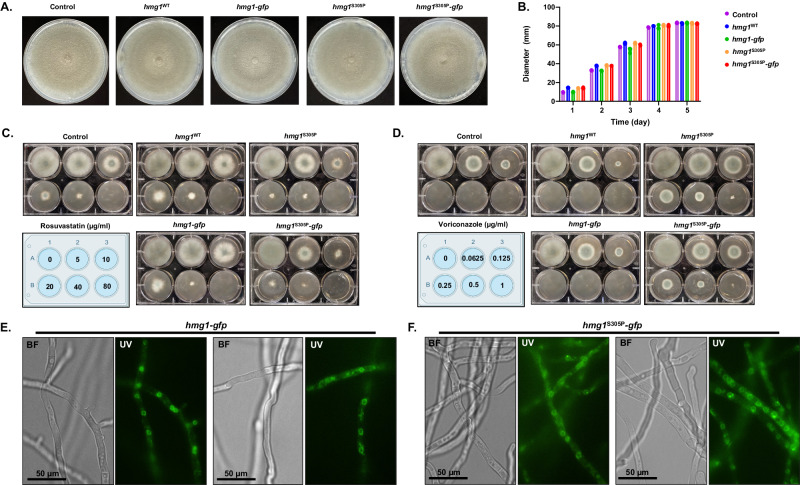
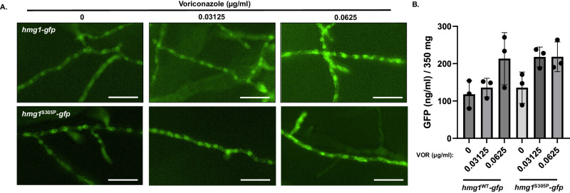
Triazole antifungals function as ergosterol biosynthesis inhibitors and are frontline therapy for invasive fungal infections, such as invasive aspergillosis. The primary mechanism of action of triazoles is through the specific inhibition of a cytochrome P450 14-α-sterol demethylase enzyme, Cyp51A/B, resulting in depletion of cellular ergosterol. Here, we uncover a clinically relevant secondary mechanism of action for triazoles within the ergosterol biosynthesis pathway. We provide evidence that triazole-mediated inhibition of Cyp51A/B activity generates sterol intermediate perturbations that are likely decoded by the sterol sensing functions of HMG-CoA reductase and Insulin-Induced Gene orthologs as increased pathway activity. This, in turn, results in negative feedback regulation of HMG-CoA reductase, the rate-limiting step of sterol biosynthesis. We also provide evidence that HMG-CoA reductase sterol sensing domain mutations previously identified as generating resistance in clinical isolates of Aspergillus fumigatus partially disrupt this triazole-induced feedback. Therefore, our data point to a secondary mechanism of action for the triazoles: induction of HMG-CoA reductase negative feedback for downregulation of ergosterol biosynthesis pathway activity. Abrogation of this feedback through acquired mutations in the HMG-CoA reductase sterol sensing domain diminishes triazole antifungal activity against fungal pathogens and underpins HMG-CoA reductase-mediated resistance.


Conflict of interest statement

The authors declare no competing interests.

Figures

Fig. 1
Fig. 1. Variant analysis of the hmg1 allele across clinical isolates identifies mutations associated with triazole resistance.
Mutations residing within the predicted sterol sensing domain (SSD) are highlighted in the green box. Triazole susceptibility phenotype, cyp51A genotype, phylogenetic relationships, and clade grouping of the clinical isolate set are indicated.
Fig. 2
Fig. 2. Hmg1-mediated triazole resistance is uniquely driven by mutations in the predicted sterol sensing domain.
A Schematic of the protein domain structure of A. fumigatus Hmg1. The location of mutations identified in genome sequences selected for this study are indicated. The previously characterized S305P mutation is indicated for context. Illustration prepared using BioRender.com. Susceptibility profiles of the parental control strain (ΔakuB-pyrG+), the manipulation control strain (hmg1WT), and the indicated hmg1 mutants to mold-active triazoles are shown for intraconazole (B), posaconazole (C), voriconazole (D), and isavuconazole (E). CLSI-based minimum inhibitory concentration (MIC) assays were performed in triplicate for each strain. Red font indicates mutant strains with shifts in susceptibility compared to controls.
Fig. 3
Fig. 3. Hmg1 SSD mutation results in altered sterol biosynthesis in response to voriconazole.
A Schematic of the mevalonate and ergosterol biosynthesis pathways in A. fumigatus. Intermediate sterols of the ergosterol pathway are indicated for their relevance to RNAseq and sterol profiling analysis results. Illustration prepared using BioRender.com. B Heat map representing differential gene expression analysis of known and predicted mevalonate and ergosterol biosynthesis pathway genes of the parental control (C), hmg1F262del (F262del), hmg1S305P (S305P), and hmg1I412S (I412S) strains. All strains were cultured in the vehicle (NT) or 0.5 X MIC voriconazole (+VOR) for 48 hours at 37 °C with shaking at 250 rpm and differentially expressed genes were determined by comparing each SSD mutant to the respective control under NT or VOR conditions. * = significantly upregulated 2-fold or more in common among the Hmg1 SSD mutant strains under NT conditions. ** = significantly upregulated 2-fold or more in common among the Hmg1 SSD mutant strains under +VOR conditions. C Relative sterol distribution in the control and Hmg1 SSD mutant strains. Strains were cultured as in B and extracted sterols analyzed by GC-MS.
Fig. 4
Fig. 4. Mutation of the Hmg1 SSD specifically alters response to triazoles and not allylamines or statins.
A Schematic of the mevalonate and ergosterol biosynthesis pathways highlighting enzymatic steps inhibited by statins (rosuvastatin), allylamines (terbinafine), and triazoles (voriconazole). Illustration prepared using BioRender.com. Radial growth assays of the parental and manipulation controls and the Hmg1 SSD mutants to assess susceptibility changes are shown for rosuvastatin (B), terbinafine (C), and voriconazole (D). Conidia (5 × 103) from each strain were spot inoculated onto RPMI media (0.2% glucose, pH 7.0) containing the indicated amount of drug and plates were cultured for 72 hours at 37 °C. Source data are provided as a Source Data file.
Fig. 5
Fig. 5. Mutations altering the transcriptional balance of insA-to-hmg1 disrupt normal response to triazole stress.
A Schematic of promoter replacement mutations for hmg1 and insA using the hspA promoter (pHspA). Illustration prepared using BioRender.com. RT-qPCR analyses to confirm overexpression is shown for hmg1 (B) and insA (C). Expression levels from two independent mutants for each gene were compared to the parental control. Averaged data represent the mean ± standard deviation. n = three biologically independent samples and statistical analyses were performed by One-Way Anova followed by an unpaired T test with significance set at 0.05 and degrees of freedom equal to 10 for individual comparisons. Radial growth assays of the parental control, hmg1 overexpression (hmg1pHspA-1 and hmg1pHspA-2), and insA overexpression (insApHspA-1 and insApHspA-2) mutants to assess susceptibility changes are shown for rosuvastatin (D), terbinafine (E), and voriconazole (F). Conidia (5 ×10³) from each strain were spot inoculated onto RPMI media (0.2% glucose, pH 7.0) containing the indicated amount of drug and plates were cultured for 72 hours at 37 °C. Source data are provided as a Source Data file.
Fig. 6
Fig. 6. Overexpression of insA restores triazole susceptibility to hmg1 overexpression mutants but does not resolve resistance due to SSD mutation.
A Schematic of promoter replacement mutations for insA in the hgm1pHspA and hmg1S305P genetic backgrounds. Illustration prepared using BioRender.com. B RT-qPCR analyses were performed in each background to ensure that the genetic manipulation at the hmg1 locus of the parental strain did not alter insA expression and to confirm that the promoter replacement resulted in overexpression of insA. Averaged data represent the mean ± standard deviation. n = three biologically independent samples and statistical analyses were performed by One-Way Anova followed by an unpaired T test with significance set at 0.05 and degrees of freedom equal to 10 for individual comparisons.Radial growth assays of the parental control, hmg1 overexpression (hmg1pHspA), hmg1/insA double overexpression (hmg1pHspA/insApHspA), hmg1 SSD (hmg1S305P), and hmg1 SSD/insA overexpression (hmg1S305P/insApHspA) mutants to assess susceptibility changes are shown for rosuvastatin (C), terbinafine (D), and voriconazole (E). Conidia (5 × 103) from each strain were spot inoculated onto RPMI media (0.2% glucose, pH 7.0) containing the indicated amount of drug and plates were cultured for 72 hours at 37 °C. Source data are provided as a Source Data file.
Fig. 7
Fig. 7. Loss of viability induced by repression of erg6 is partially blocked by Hmg1 SSD mutation.
Models for sterol intermediate-induced negative feedback of Hmg1 through promotion of InsA binding under normal conditions (A), under triazole stress (B), and under doxycycline-mediated repression of erg6 expression in an erg6pTetOff mutant (C). Illustration prepared using BioRender.com. D The parental control strains (hmg1WT and hmg1S305P) and their erg6pTetOff derivatives (hmg1WT/erg6pTetOff and hmg1S305P/erg6pTetOff, respectively) were utilized for spot dilution assays on RPMI media (0.2% glucose, pH 7.0) with the indicated concentrations of doxycycline. Conidial inoculum concentrations for each strain were (from left to right) 5 × 104, 5 × 103, 5 × 102, and 5 × 101 total conidia. Note the diminished ability of erg6 repression (i.e., increasing concentrations of exogenous doxycycline) to inhibit growth in the background of an hmg1 SSD mutant (hmg1S305P).
Fig. 8
Fig. 8. Loss of the conserved ERAD ubiquitin ligase, hrdA, does not resolve hypersusceptibility generated by insA overexpression.
A Schematic of the potential mechanisms for Hmg1 regulation involving InsA and HrdA based on the known mechanisms in mammalian systems (i), the budding yeast Saccharomyces cerevisiae (ii) and the fission yeast Schizosaccharomyces pombe (iii). Illustration prepared using BioRender.com. B Voriconazole susceptibility assays using the parental control, hrdA deletion strain (ΔhrdA), insA overexpression strain (insApHspA), and the insA overexpression/hrdA deletion double mutant (insApHspAhrdA). Conidia (5 × 103) from each strain were spot inoculated onto RPMI media (0.2% glucose, pH 7.0) containing the indicated amount of drug and plates were cultured for 72 hours at 37 °C. Source data are provided as a Source Data file.
Fig. 9
Fig. 9. Hmg1 SSD mutations do not alter localization of Hmg1 to the A. fumigatus endoplasmic reticulum.
A Colony morphology comparison of the control, Hmg1 parental (hmg1WT and hmg1S305P), and Hmg1-GFP strains (hmg1WT-gfp and hmg1S305P-gfp) after 5 days growth on minimal media. B Quantitation of colony diameter for the indicated strains at days 1, 2, 3, 4, and 5 post-inoculation. Assays were completed in biological triplicate. C Rosuvastatin and D voriconazole susceptibility assays for the control, Hmg1 parental, and Hmg1-GFP strains. Assays were completed as described in Figure X. Drug concentrations for each well are noted in the included plate diagrams. E Hmg1WT-GFP and F Hmg1S305P-GFP localization analyzed by epifluorescence microscopy. Conidia from each strain were inoculated into minimal media, incubated for 12 hrs at 37 °C, and then mounted for microcopy. Images were captured using an exposure time of 600 ms. Note localization to regularly spaced ring structures indicative of A. fumigatus peri-nuclear endoplasmic reticulum. BF = brightfield; UV = ultraviolet. Microscopy experiments were completed three times independently with similar results. Source data are provided as a Source Data file.
Fig. 10
Fig. 10. Triazole stress does not reduce Hmg1 protein abundance.
A Localization analyses of Hmg1-GFP in response to sub-lethal triazole stress in the hmg1WT-gfp and hmg1S305P-gfp strains. All images were captured with an exposure time of 600 ms. Note apparent increase in intensity and mislocalization of the GFP fluorescent signal at 0.0625 µg/ml voriconazole. Scale bar = 50 µm. Microscopy experiments were completed three times independently with similar results. B Quantitation of GFP in lysates of the hmg1WT-gfp and hmg1S305P-gfp strains cultured for 12 hrs in the presence of 0, 0.03125, or 0.0625 µg/ml voriconazole. Data are presented as the mean ± standard deviation. Assays were performed in biological triplicate and statistical analyses completed using one-way ANOVA and Tukey’s Test post-hoc. Despite a trend for increased GFP abundance in the presence of voriconazole, no significant differences were noted among the groups. Source data are provided as a Source Data file.

References

    1. Bongomin F, Gago S, Oladele RO, Denning DW. Global and multi-national prevalence of fungal diseases—estimate precision. J. Fungi. 2017;3:57. doi: 10.3390/jof3040057. - DOI - PMC - PubMed
    1. Parker JE, et al. Resistance to antifungals that target CYP51. J. Chem. Biol. 2014;7:143–161. doi: 10.1007/s12154-014-0121-1. - DOI - PMC - PubMed
    1. Patterson TF, et al. Practice Guidelines for the Diagnosis and Management of Aspergillosis: 2016 Update by the Infectious Diseases Society of America. Clin. Infect. Dis. 2016;63:e1–e60. doi: 10.1093/cid/ciw326. - DOI - PMC - PubMed
    1. Lestrade PP, et al. Voriconazole resistance and mortality in invasive aspergillosis: a multicenter retrospective cohort study. Clin. Infect. Dis. 2018;68:1463–1471. doi: 10.1093/cid/ciy859. - DOI - PubMed
    1. Baddley JW, et al. Patterns of susceptibility of Aspergillus isolates recovered from patients enrolled in the transplant-associated infection surveillance network. J. Clin. Microbiol. 2009;47:3271–3275. doi: 10.1128/JCM.00854-09. - DOI - PMC - PubMed

Publication types

MeSH terms