. 2024 Apr 29;14(1):9846.
doi: 10.1038/s41598-024-59773-2.

Machine learning approach for recognition and morphological analysis of isolated astrocytes in phase contrast microscopy

Affiliations

Machine learning approach for recognition and morphological analysis of isolated astrocytes in phase contrast microscopy

Egor V Yakovlev et al. Sci Rep. .

Abstract

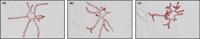
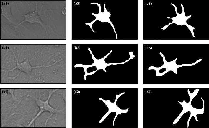
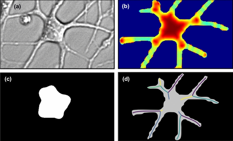
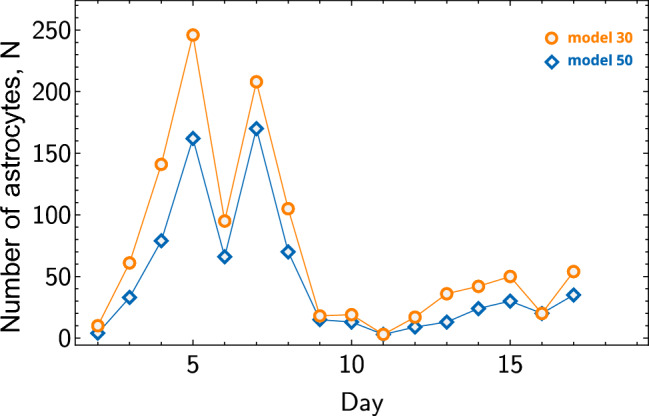
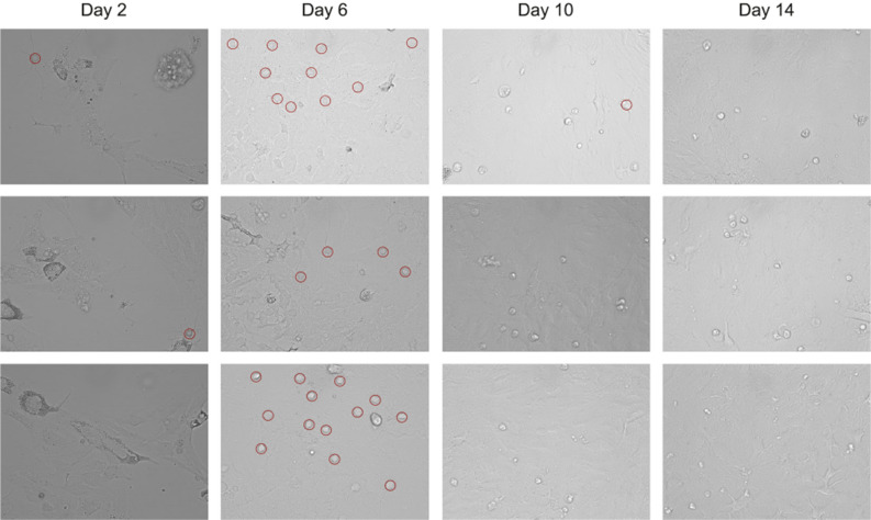
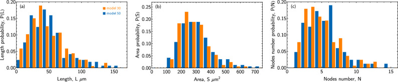
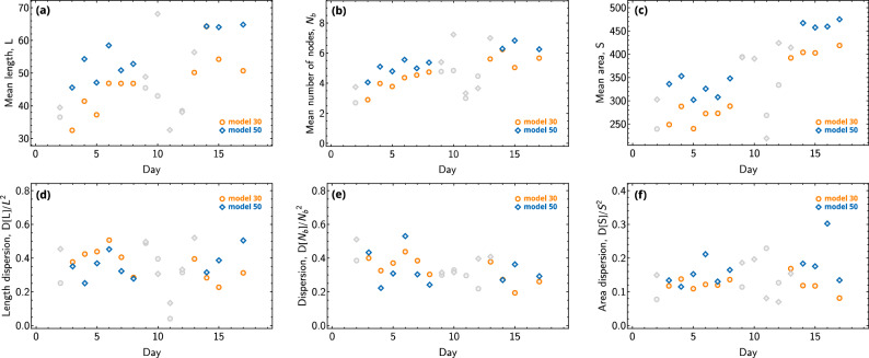
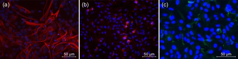
Astrocytes are glycolytically active cells in the central nervous system playing a crucial role in various brain processes from homeostasis to neurotransmission. Astrocytes possess a complex branched morphology, frequently examined by fluorescent microscopy. However, staining and fixation may impact the properties of astrocytes, thereby affecting the accuracy of the experimental data of astrocytes dynamics and morphology. On the other hand, phase contrast microscopy can be used to study astrocytes morphology without affecting them, but the post-processing of the resulting low-contrast images is challenging. The main result of this work is a novel approach for recognition and morphological analysis of unstained astrocytes based on machine-learning recognition of microscopic images. We conducted a series of experiments involving the cultivation of isolated astrocytes from the rat brain cortex followed by microscopy. Using the proposed approach, we tracked the temporal evolution of the average total length of branches, branching, and area per astrocyte in our experiments. We believe that the proposed approach and the obtained experimental data will be of interest and benefit to the scientific communities in cell biology, biophysics, and machine learning.


Conflict of interest statement

The authors declare no competing interests.

Figures

Figure 1
Figure 1
Example of a labeled image of an astrocyte from the rat brain cortex obtained with phase contrast microscopy in our experiments.
Figure 2
Figure 2
The region of interest (ROI) in the detection of the morphology of an astrocyte. The first column represents the image of an astrocyte, the second column represents the detection by the neural network Model 30, and the third column represents the detection by the neural network Model 50.
Figure 3
Figure 3
False positive (FP) results of astrocyte detection by Model 30. The segmented mask is highlighted in red.
Figure 4
Figure 4
Segmentation of the soma in an astrocyte. The (a) is the original microscope image, the (b) is the image colored according to the directional ratio, the (c) is the segmented soma and the (d) is the segmented mask of an astrocyte and its skeletonized branches, different colors correspond to the recognized segments.
Figure 5
Figure 5
Number of detected astrocytes. Blue and orange symbols correspond to Models 50 and 30, respectively, with lines added for better visibility.
Figure 6
Figure 6
Images of astrocytes recorded on 2nd, 6th, 10th, 14th day respectively. The images represent a random set from microscopic data taken on the respective days. The red circles indicate the astrocytes that fell within the imaged area.
Figure 7
Figure 7
The histograms of astrocyte parameters, as measured on the 7th day of experiments. (a) The distribution by lengths, (b) the distribution by areas, and (c) the distribution by the number of nodes. The orange bars represent the results of Model 30, while the blue bars represent the results of Model 50. The touching blue and red bars refer to the same value.
Figure 8
Figure 8
The temporal dependencies of astrocyte parameters are shown: (a) illustrates the average length of branches L, (b) shows the average number of nodes per astrocyte Nb, and (c) presents the average area S of the astrocyte. (df) The dependencies of dispersion of the considered characteristics, normalized to the mean value squared, D[L]/L2, D[Nb]/Nb2, and D[S]/S2 respectively. The orange symbols represent the results of Model 30, while blue symbols represent the results of Model 50. Data points calculated from samples of fewer than 20 astrocytes are colored gray.
Figure 9
Figure 9
Identification of culture cells: (a) astrocytes in the primary culture: the cell bodies and processes are stained with GFAP antibody (red), the nuclei are stained with DAPI (blue), (b) microglia in the primary culture: cell bodies and processes are stained with Iba1 antibody (red), the nuclei are stained with DAPI (blue), (c) pericytes in the primary culture: the cell bodies and processes are stained with PDGFRB antibody (green), the nuclei are stained with DAPI (blue).
Figure 10
Figure 10
Images acquired during the same experiment (on the same day and on an identical setup) to check for possible bias between experiments under nominally identical conditions.

References

    1. Cabezas R, et al. Growth factors and astrocytes metabolism: Possible roles for platelet derived growth factor. Med. Chem. 2016;12:204–210. doi: 10.2174/1573406411666151019120444. - DOI - PubMed
    1. Nag, S. & Walker, J. The Blood–Brain and Other Neural Barriers: Reviews and Protocols (Humana Press, 2011).
    1. Siracusa R, Fusco R, Cuzzocrea S. Astrocytes: Role and functions in brain pathologies. Front. Pharmacol. 2019 doi: 10.3389/fphar.2019.01114. - DOI - PMC - PubMed
    1. Freeman MR. Specification and morphogenesis of astrocytes. Science. 2010;330:774–778. doi: 10.1126/science.1190928. - DOI - PMC - PubMed
    1. Guillamon-Vivancos T, Gomez-Pinedo U, Matias-Guiu J. Astrocytes in neurodegenerative diseases (I): Function and molecular description. Neurologia (English Edition) 2015;30:119–129. doi: 10.1016/j.nrleng.2014.12.005. - DOI - PubMed

Publication types