. 2024 Aug;45(8):1715-1726.
doi: 10.1038/s41401-024-01258-z. Epub 2024 Apr 29.

Baicalein triggers ferroptosis in colorectal cancer cells via blocking the JAK2/STAT3/GPX4 axis

Affiliations

Baicalein triggers ferroptosis in colorectal cancer cells via blocking the JAK2/STAT3/GPX4 axis

Jian-Qin Lai et al. Acta Pharmacol Sin. 2024 Aug.

Abstract

Colorectal cancer (CRC) is a prevalent form of gastrointestinal malignancy with challenges in chemotherapy resistance and side effects. Effective and low toxic drugs for CRC treatment are urgently needed. Ferroptosis is a novel mode of cell death, which has garnered attention for its therapeutic potential against cancer. Baicalein (5, 6, 7-trihydroxyflavone) is the primary flavone extracted from the dried roots of Scutellaria baicalensis that exhibits anticancer effects against several malignancies including CRC. In this study, we investigated whether baicalein induced ferroptosis in CRC cells. We showed that baicalein (1-64 μM) dose-dependently inhibited the viability of human CRC lines HCT116 and DLD1. Co-treatment with the ferroptosis inhibitor liproxstatin-1 (1 μM) significantly mitigated baicalein-induced CRC cell death, whereas autophagy inhibitor chloroquine (25 μM), necroptosis inhibitor necrostatin-1 (10 μM), or pan-caspase inhibitor Z-VAD-FMK (10 μM) did not rescue baicalein-induced CRC cell death. RNA-seq analysis confirmed that the inhibitory effect of baicalein on CRC cells is associated with ferroptosis induction. We revealed that baicalein (7.5-30 μM) dose-dependently decreased the expression levels of GPX4, key regulator of ferroptosis, in HCT116 and DLD1 cells by blocking janus kinase 2 (JAK2)/STAT3 signaling pathway via direct interaction with JAK2, ultimately leading to ferroptosis in CRC cells. In a CRC xenograft mouse model, administration of baicalein (10, 20 mg/kg, i.g., every two days for two weeks) dose-dependently inhibited the tumor growth with significant ferroptosis induced by inhibiting the JAK2/STAT3/GPX4 axis in tumor tissue. This study demonstrates that ferroptosis contributes to baicalein-induced anti-CRC activity through blockade of the JAK2/STAT3/GPX4 signaling pathway, which provides evidence for the therapeutic application of baicalein against CRC.

Keywords: GPX4; JAK2/STAT3; baicalein; colorectal cancer; ferroptosis; liproxstatin-1.


Conflict of interest statement

The authors declare no competing interests.

Figures
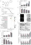
Fig. 1
Fig. 1. Ferroptosis induction is associated with anti-CRC effect of baicalein.
a Chemical structure of baicalein (Bai). b Cell viability of DLD1, HCT116, and NCM460 cells treated with baicalein was examined by the CCK8 assay. c KEGG pathway enrichment analysis was performed in HCT116 cells treated by baicalein (30 μM). d Following treatment with baicalein for 24 h, ROS levels were analyzed by flow cytometer. GSH (e) and MDA (f) assay were performed after baicalein treatment in DLD1 and HCT116 cells. g MMP was detected and quantified by flow cytometry with mitoscreen JC-1 staining (ratio of red JC-1 aggregates to green JC-1 monomers). h FerroOrange fluorescent probe was used to image Fe2+ in HCT116 cells treated with or without baicalein (30 μM). Blue, DAPI-stained nuclei; yellow, Fe2+. Scale bar, 50 μm. i Transmission Electron microscope was used to observe the cell structure after baicalein (30 μM) treatment for 24 h. Black arrow, mitochondrial morphology in untreated cells; red arrow, mitochondrial morphology in baicalein-treated cells. Scale bar, 500 nm. j Expression of ferroptosis-related proteins in DLD1 and HCT116 cells by Western blotting. Data are presented as the mean ± SD, n = 3, *P < 0.05, **P < 0.01, ***P < 0.001, compared with control group.
Fig. 2
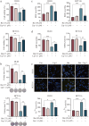
Fig. 2. Baicalein-mediated cytotoxicity is dependent on ferroptosis.
a Cell viability assessment in DLD1 and HCT116 cells treated with or without Lip-1 (1 μM) in the presence of baicalein. b Colony formation assay and quantitative analysis in DLD1 and HCT116 cells treated with or without Lip-1 in the presence of baicalein. c Analysis of ROS levels in DLD1 and HCT116 cells treated with or without Lip-1 in the presence of baicalein. d GSH levels in DLD1 and HCT116 cells treated with or without Lip-1 in the presence of baicalein. e Detection of Fe2+ levels using the FerroOrange fluorescent probe in DLD1 and HCT116 cells treated with or without Lip-1 in the presence of baicalein. Blue represents DAPI-stained nuclei, and yellow represents Fe2+. Scale bar, 50 μm. Data are presented as the mean ± SD, n = 3, *P < 0.05, **P < 0.01, ***P < 0.001.
Fig. 3
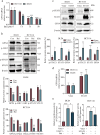
Fig. 3. Baicalein blocks JAK2/STAT3/GPX4 axis for ferroptosis induction.
a qRT–PCR analysis of GPX4 mRNA expression in DLD1 and HCT116 cells treated with various concentrations of baicalein. b Western blotting analysis of JAK2, p-JAK2, STAT3, p-STAT3, and GPX4 protein levels in cells treated with or without baicalein (30 μM). c, d Western blotting (c) and qRT-PCR (d) analyses of changes in GPX4 expression induced by STAT3 in DLD1 and HCT116 cells. e Luciferase reporter assays in DLD1 and HCT116 cells overexpressing STAT3 and GPX4 reporter plasmids treated with or without baicalein (30 μM). Data are presented as the mean ± SD, n = 3, *P < 0.05, **P < 0.01, ***P < 0.001.
Fig. 4
Fig. 4. Baicalein promotes ferroptosis by targeting JAK2.
Western blotting (a) and qRT–PCR (b) analysis of STAT3, p-STAT3, and GPX4 levels in CRC cells subjected to indicated treatment (Bai 30 μM). c Cell viability assessed by CCK-8 assay at 0, 24, 48, and 72 h in CRC cells from different treatment groups (Bai 7.5 μM). GSH (d) and MDA (e) levels in CRC cells from the indicated treatments (Bai 30 μM). f Molecular docking model illustrating the binding of baicalein to JAK2. g CETSA showed JAK2 target engagement by baicalein in HCT116 cells. h Effect of baicalein on JAK2 kinase activity. i Representative data from ITC indicate the binding of baicalein and JAK2. Data are presented as the mean ± SD, n = 3, *P < 0.05, **P < 0.01, ***P < 0.001.
Fig. 5
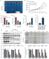
Fig. 5. Baicalein triggers ferroptosis in CRC xenograft model.
a Representative images of tumor samples from each experimental group. b Tumor volume in each group. c Analysis of tumor growth inhibition in each group. d, e Levels of GSH (d) and MDA (e) in tumor tissues. f Detection of free iron deposition in tumor tissue using the Prussian Blue Iron Stain Kit. Scale bar, 100 μm. g Western blotting analysis of JAK2, p-JAK2, STAT3, p-STAT3, and GPX4 protein levels in tumors. h The expression of JAK2, p-JAK2, STA3, p-STAT3, GPX4, and Ki-67 in tumor tissues was determined by immunohistochemical staining. Scale bar, 50 μm. Data are expressed as the mean ± SD, n = 3, *P < 0.05, **P < 0.01, ***P < 0.001, compared with vehicle group.
Fig. 6
Fig. 6. Schematic illustration of baicalein-induced ferroptosis via JAK2/STAT3/GPX4 axis in CRC cells.
In CRC cells, baicalein directly interacts with JAK2, blocks the JAK2/STAT3 signaling pathway, leading to downregulation of GPX4, subsequently promoting ROS production, lipid peroxidation, ultimately inducing ferroptosis.

References

    1. Sung H, Ferlay J, Siegel RL, Laversanne M, Soerjomataram I, Jemal A, et al. Global Cancer Statistics 2020: GLOBOCAN Estimates of Incidence and Mortality Worldwide for 36 Cancers in 185 Countries. CA Cancer J Clin. 2021;71:209–49. 10.3322/caac.21660 - DOI - PubMed
    1. García-Alfonso P, Torres G, García G, Gallego I, Ortega L, Sandoval C, et al. FOLFOXIRI plus biologics in advanced colorectal cancer. Expert Opin Biol Ther. 2019;19:411–22. 10.1080/14712598.2019.1595580 - DOI - PubMed
    1. Haraldsdottir S, Einarsdottir HM, Smaradottir A, Gunnlaugsson A, Halfdanarson TR. Colorectal cancer - review. Laeknabladid. 2014;100:75–82. - PubMed
    1. Biller LH, Schrag D. Diagnosis and treatment of metastatic colorectal cancer: a review. JAMA. 2021;325:669–85. 10.1001/jama.2021.0106 - DOI - PubMed
    1. Brody H. Colorectal cancer. Nature. 2015;521:S1. 10.1038/521S1a - DOI - PubMed

MeSH terms