

Artificial intelligence assists identification and pathologic classification of glomerular lesions in patients with diabetic nephropathy
- PMID: 38684996
- PMCID: PMC11059590
- DOI: 10.1186/s12967-024-05221-8
Artificial intelligence assists identification and pathologic classification of glomerular lesions in patients with diabetic nephropathy
Abstract
Background: Glomerular lesions are the main injuries of diabetic nephropathy (DN) and are used as a crucial index for pathologic classification. Manual quantification of these morphologic features currently used is semi-quantitative and time-consuming. Automatically quantifying glomerular morphologic features is urgently needed.
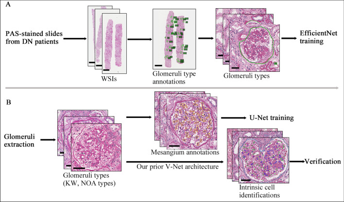
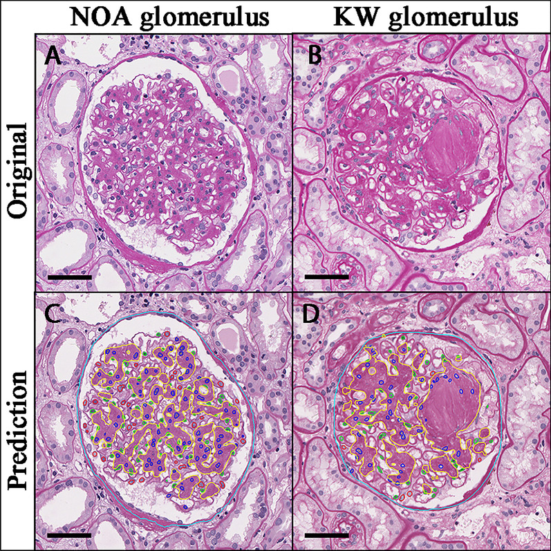
Methods: A series of convolutional neural networks (CNN) were designed to identify and classify glomerular morphologic features in DN patients. Associations of these digital features with pathologic classification and prognosis were further analyzed.
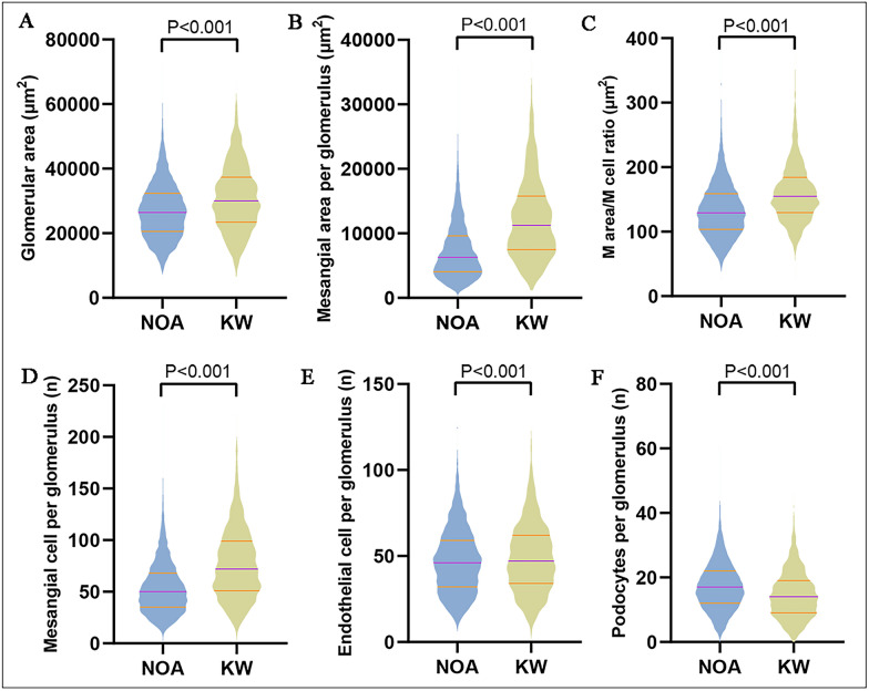
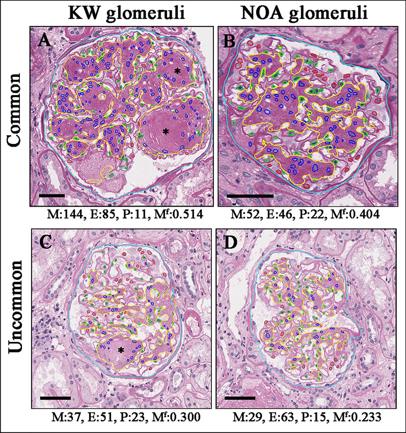
Results: Our CNN-based model achieved a 0.928 F1-score for global glomerulosclerosis and 0.953 F1-score for Kimmelstiel-Wilson lesion, further obtained a dice of 0.870 for the mesangial area and F1-score beyond 0.839 for three glomerular intrinsic cells. As the pathologic classes increased, mesangial cell numbers and mesangial area increased, and podocyte numbers decreased (p for all < 0.001), while endothelial cell numbers remained stable (p = 0.431). Glomeruli with Kimmelstiel-Wilson lesion showed more severe podocyte deletion compared to those without (p < 0.001). Furthermore, CNN-based classifications showed moderate agreement with pathologists-based classification, the kappa value between the CNN model 3 and pathologists reached 0.624 (ranging from 0.529 to 0.688, p < 0.001). Notably, CNN-based classifications obtained equivalent performance to pathologists-based classifications on predicting baseline and long-term renal function.
Conclusion: Our CNN-based model is promising in assisting the identification and pathologic classification of glomerular lesions in DN patients.
Keywords: Artificial intelligence; Diabetic nephropathy; Glomerulus; Identification; Pathology.
© 2024. The Author(s).
Conflict of interest statement
All authors declare that no potential conflict of interest exists.
Figures
Similar articles
-
Pathologic classification of diabetic nephropathy.J Am Soc Nephrol. 2010 Apr;21(4):556-63. doi: 10.1681/ASN.2010010010. Epub 2010 Feb 18. J Am Soc Nephrol. 2010. PMID: 20167701
-
Renal pathology patterns in type II diabetes mellitus: relationship with retinopathy. The Collaborative Study Group.Nephrol Dial Transplant. 1998 Oct;13(10):2547-52. doi: 10.1093/ndt/13.10.2547. Nephrol Dial Transplant. 1998. PMID: 9794557 Clinical Trial.
-
Computational Segmentation and Classification of Diabetic Glomerulosclerosis.J Am Soc Nephrol. 2019 Oct;30(10):1953-1967. doi: 10.1681/ASN.2018121259. Epub 2019 Sep 5. J Am Soc Nephrol. 2019. PMID: 31488606 Free PMC article.
-
Pathology of human diabetic nephropathy.Contrib Nephrol. 2011;170:36-47. doi: 10.1159/000324942. Epub 2011 Jun 9. Contrib Nephrol. 2011. PMID: 21659756 Review.
-
Renal morphologic lesions reminiscent of diabetic nephropathy.Arch Pathol Lab Med. 2013 Mar;137(3):351-9. doi: 10.5858/arpa.2012-0243-RA. Arch Pathol Lab Med. 2013. PMID: 23451746 Review.
Cited by
-
Artificial Intelligence Models in Diagnosis and Treatment of Kidney Diseases: Current Status and Prospects.Kidney Dis (Basel). 2025 Jun 12;11(1):491-507. doi: 10.1159/000546397. eCollection 2025 Jan-Dec. Kidney Dis (Basel). 2025. PMID: 40672962 Free PMC article.
-
Multimodal deep learning improving the accuracy of pathological diagnoses for membranous nephropathy.Ren Fail. 2025 Dec;47(1):2528106. doi: 10.1080/0886022X.2025.2528106. Epub 2025 Jul 14. Ren Fail. 2025. PMID: 40659521 Free PMC article.
-
GlomSAM: Hybrid customized SAM for multi-glomerular detection and segmentation in immunofluorescence images.PLoS One. 2025 Apr 14;20(4):e0321096. doi: 10.1371/journal.pone.0321096. eCollection 2025. PLoS One. 2025. PMID: 40228213 Free PMC article.
References
-
- Fiorentino M, Bolignano D, Tesar V, et al. Renal biopsy in patients with diabetes: a pooled meta-analysis of 48 studies. Nephrol Dial Transplant. 2017;32:97–110. - PubMed
Publication types
MeSH terms
Grants and funding
LinkOut - more resources
Full Text Sources
Medical
Research Materials
