. 2024 Apr 29;43(1):128.
doi: 10.1186/s13046-024-03024-9.

Blocking the MIF-CD74 axis augments radiotherapy efficacy for brain metastasis in NSCLC via synergistically promoting microglia M1 polarization

Affiliations

Blocking the MIF-CD74 axis augments radiotherapy efficacy for brain metastasis in NSCLC via synergistically promoting microglia M1 polarization

Lichao Liu et al. J Exp Clin Cancer Res. .

Abstract

Background: Brain metastasis is one of the main causes of recurrence and death in non-small cell lung cancer (NSCLC). Although radiotherapy is the main local therapy for brain metastasis, it is inevitable that some cancer cells become resistant to radiation. Microglia, as macrophages colonized in the brain, play an important role in the tumor microenvironment. Radiotherapy could activate microglia to polarize into both the M1 and M2 phenotypes. Therefore, searching for crosstalk molecules within the microenvironment that can specifically regulate the polarization of microglia is a potential strategy for improving radiation resistance.

Methods: We used databases to detect the expression of MIF in NSCLC and its relationship with prognosis. We analyzed the effects of targeted blockade of the MIF/CD74 axis on the polarization and function of microglia during radiotherapy using flow cytometry. The mouse model of brain metastasis was used to assess the effect of targeted blockade of MIF/CD74 axis on the growth of brain metastasis.

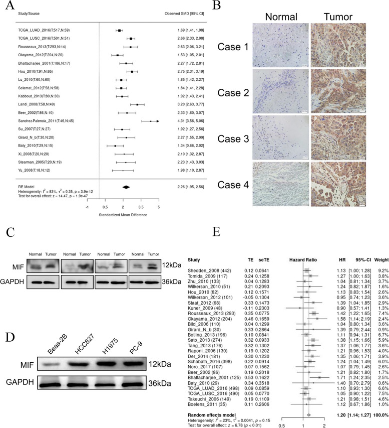
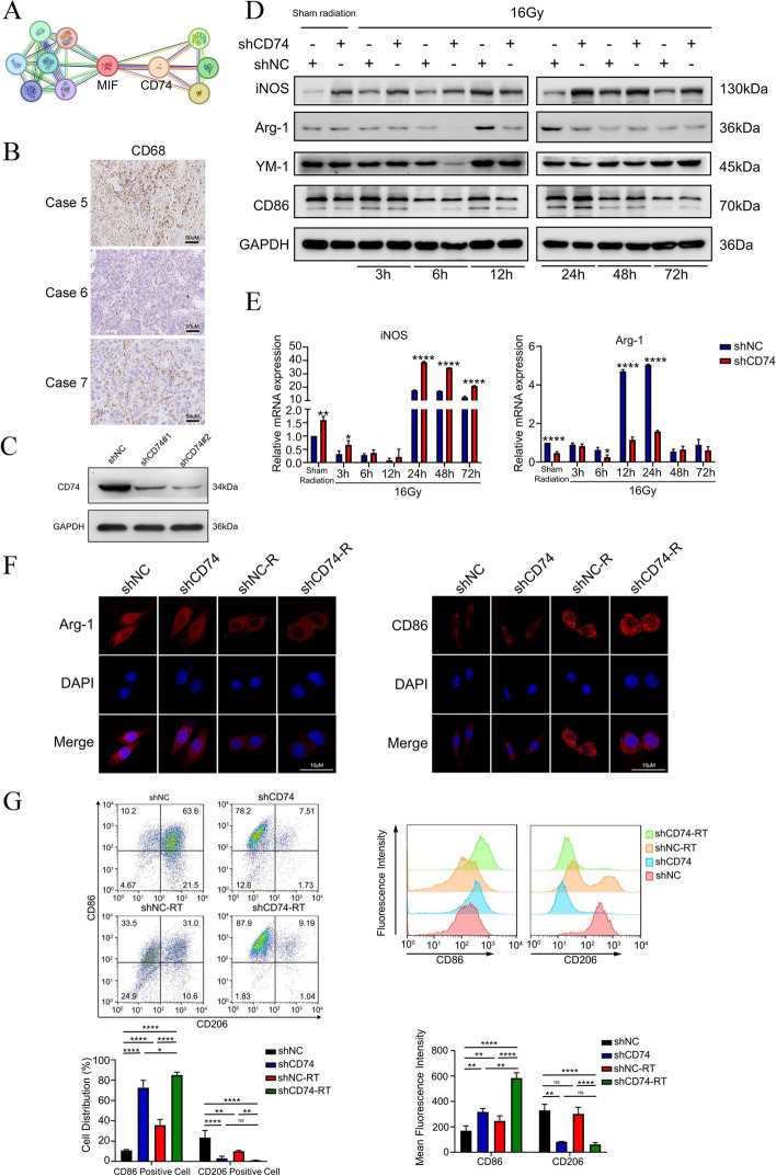
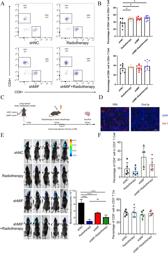
Result: Our findings reveals that the macrophage migration inhibitory factor (MIF) was highly expressed in NSCLC and is associated with the prognosis of NSCLC. Mechanistically, we demonstrated CD74 inhibition reversed radiation-induced AKT phosphorylation in microglia and promoted the M1 polarization in combination of radiation. Additionally, blocking the MIF-CD74 interaction between NSCLC and microglia promoted microglia M1 polarization. Furthermore, radiation improved tumor hypoxia to decrease HIF-1α dependent MIF secretion by NSCLC. MIF inhibition enhanced radiosensitivity for brain metastasis via synergistically promoting microglia M1 polarization in vivo.

Conclusions: Our study revealed that targeting the MIF-CD74 axis promoted microglia M1 polarization and synergized with radiotherapy for brain metastasis in NSCLC.

Keywords: Brain metastases; CD74; MIF; Microglia; NSCLC; Radiotherapy.


Conflict of interest statement

The authors declare that they have no competing interests.

Figures

Fig. 1
Fig. 1
MIF was elevated in NSCLC and indicated a poor prognosis a. Meta analysis of MIF expression in tumor tissue and normal tissues in LUNG CANCER EXPLORER website. b IHC staining of MIF in NSCLC brain metastatic tissue and paired paracancerous tissue c. Difference of MIF protein level in NSCLC brain metastases and paired paracancerous surgical tissues. d. Difference of MIF protein level in Beas-2B, HCC827, H1975, and PC-9 cells. e. Survival meta-analysis of correlation between the expression of MIF and survival of NSCLC patient in LUNG CANCER EXPLORER website
Fig. 2
Fig. 2
Inhibiting CD74 in microglia in combination with radiation synergistically promoted M1 polarization a. Molecules interacting with MIF in STRING database. b. IHC staining of CD86 in NSCLC brain metastatic tissue. c. Western blotting of CD74 in BV2 cells transduced with either shNC or shCD74 Lentivirus. d. Western blotting for the expression of M1 marker iNOS, CD86 and M2 marker Ym-1, Arg-1 in shNC-BV2 cell or shCD74-BV2 cell in sham radiation and 3, 6, 12, 24, 48, and 72 h after 16-Gy radiation. e. Total RNA of shNC-BV2 cell or shCD74-BV2 cell was extracted in sham radiation and 3, 6, 12, 24, 48, and 72 h after radiation, and mRNA level of iNOS and Arg-1 were analyzed by real-time qPCR (n = 3). f. Representative microphotographs of immunofluorescence staining showing expression of Arg-1(left) and CD86(right) in shNC-BV2 cell or shCD74-BV2 cell with or without 16 Gy radiation at 6 h after radiation. g. The ratio of CD86 positive and CD206 positive BV-2 cell was analyzed by flow cytometry and the data was processed with FlowJo (version 10.0) program (left). The mean fluorescence intensity of CD86 and CD206 in BV-2 cell was analyzed by flow cytometry and the data was processed with FlowJo (version 10.0) program (right), n = 3
Fig. 3
Fig. 3
CD74 inhibition reversed radiation-induced AKT phosphorylation in microglia and promoted the M1 polarization in combination of radiation a. Western blotting was used for analyzing the change of phosphorylated-AKT(p-AKT) and AKT in shNC-BV2 cell or shCD74-BV2 cell in sham radiation and 3, 6, 12, 24, 48, and 72 h after 16 Gy radiation. b. Western blotting for the change of phosphorylated-ERK1/2(p-ERK1/2) and ERK1/2 in shNC-BV2 cell or shCD74-BV2 cell in sham radiation and 3, 6, 12, 24, 48, and 72 h after 16 Gy radiation. c. Western blotting for the change of phosphorylated-AKT(p-AKT), AKT, YM-1, Arg-1, iNOS and CD86 in BV2 cell with or without LY294002 in sham radiation and 3, 6, 12, 24, 48, and 72 h after 16-Gy radiation. d. Total RNA of BV2 cell was extracted in sham radiation or 16-Gy radiation at 6 h after radiation with or without LY294002, and mRNA level of iNOS, Arg-1, CD86 and YM-1 were analyzed by real-time PCR (n = 3). e. Representative microphotographs of immunofluorescence staining showing level of F4/80(green)/CD86(red, left) and F4/80(green)/Arg-1(red, right) in BV-2 cultured with or without LY294002 plus 16 Gy radiation at 6 h after radiation. f. Mean fluorescence intensity of CD86 and CD206 in BV-2 cell was analyzed by flow cytometry(left) and the data was processed with FlowJo (version 10.0) program (right, n = 3). g. Mean fluorescence intensity of BV2 cell phagocytic microspheres analyzed by flow cytometry (n = 3). h. The phagocytic activity of BV2 is defined as the level of green microspheres engulfed by the red phalloidin labelled-BV2. Representative fluorescent images showing phagocytic activity of BV-2 cultured with or without LY294002 plus 16 Gy radiation at 6 h after radiation. i. Immunofluorescence was used to analyze the apoptosis rate of BV2 cells (n = 3)
Fig. 4
Fig. 4
Blocking the MIF-CD74 interaction between NSCLC and microglia promoted microglia M1 polarization a. Co-culture sketch map of CD74-shRNA lentivirus transduced BV2 cells and MIF-shRNA lentivirus transduced Lewis cells. After 16-Gy radiation, BV2 cell were immediately co cultured with Lewis cells for 24 h. b. Total RNA of BV2 cell was extracted after 24 h co-culture, and mRNA expressions of iNOS, Arg-1, CD86 and YM-1 were analyzed by real-time PCR (n = 3). c. Western blotting for the expression of iNOS, CD86, Arg-1 and YM-1 in BV2 cell after 24 h co-culture. d. Representative fluorescent images showing different phagocytosis of microspheres (green) with phalloidin (red) in BV-2 after 24 h co-culture. e. Representative microphotographs of immunofluorescence staining showing expression of Arg-1(left) and CD86(right) in BV-2 cell after 24 h co-culture. f. The ratio of CD86 positive and CD206 positive BV-2 cell was analyzed by flow cytometry and the data was processed with FlowJo (version 10.0) program (n = 3). g. The mean fluorescence intensity of CD86 and CD206 in BV-2 cell was analyzed by flow cytometry and the data was processed with FlowJo (version 10.0) program (n = 3)
Fig. 5
Fig. 5
Radiation improved tumor hypoxia and impeded HIF-1α-dependent MIF secretion by NSCLC. a Representative microphotographs of immunofluorescence staining showing expression of CD34(red) and α-SMA (green) in shNC or shMIF BM mouse model with or without 10 Gy radiation therapy (n = 3). b. IHC staining of HIF-1α in brain metastatic tissue of BM mouse model. c. Correlation analysis of MIF and HIF-1α in NSCLC patients in R2 database. d. Dose–response curves of KC7F2 in Lewis cell. e. Western blotting for the level of HIF-1α and MIF in Lewis cell under 1% O2 with 0 μM, 10μ, 20 μM, 30 μM, 60 μM KC7F2. f. Western blotting for the expression of HIF-1α and MIF in Lewis cell under 1% O2 after 3 h, 6 h, 12 h, 24 h, 48 h. g. Western blotting for the expression of HIF-1α and MIF in Lewis cell under 1% O2 without KC7F2 and with 30 μM KC7F2 after 3 h, 6 h, 12 h, 24 h, 48 h. h. Change of MIF concentration in Lewis cell’s supernatant under 21% or 1% O2 with or without KC7F2 (n = 3). i. Binding ability of HIF-1α and MIF promoter in EMSA assay. j. Western blotting for the expression of iNOS, CD86, Arg-1 and YM-1 in shNC or shCD74 BV2 cell of the co-culture system. 30 μM KC7F2 was added to the culture medium of Lewis cell in the co-culture system. k. Representative microphotographs of immunofluorescence staining showing expression of Arg-1 and CD86 in shNC or shCD74 BV2 cell of the co-culture system. 30 μM KC7F2 was added to the culture medium of Lewis cell in the co-culture system
Fig. 6
Fig. 6
MIF inhibition enhanced radiosensitivity for brain metastasis via synergistically promoting microglia M1 polarization in vivo a. The schema for animal studies. b. Representative bioluminescent imaging of BM model assay with shNC and shMIF Luc-Lewis cells carotid artery injected in C57BL/6 mice with or without 10 Gy radiotherapy(n = 10). c. HE staining of BM mice model assay. d. Survival analysis for BM mice model (n = 5). e. Body weight measurement for BM mice model (n = 5). f. Western blotting for the expression of iNOS and Arg-1 in brain metastasis of BM model. g. Immunofluorescence staining of brain metastasis tissue of BM mice model. h. At 7 days after radiotherapy, the ratio of CD86 positive and CD206 positive BV-2 cell was analyzed by flow cytometry and the data was processed with FlowJo (version 10.0) program (left). The mean fluorescence intensity of CD86 and CD206 in BV-2 cell was analyzed by flow cytometry and the data was processed with FlowJo (version 10.0) program(right, n = 3). i. At 14 days after radiotherapy, the ratio of CD86 positive and CD206 positive BV-2 cell was analyzed by flow cytometry and the data was processed with FlowJo (version 10.0) program (left). The mean fluorescence intensity of CD86 and CD206 in BV-2 cell was analyzed by flow cytometry and the data was processed with FlowJo(version 10.0) program(right, n = 3)
Fig. 7
Fig. 7
The synergistic antitumor effect of MIF inhibition and radiotherapy was dependent on macrophages a. Representative Flowchart in shNC or shMIF brain metastasis tissue of BM mice model. b Percentage of CD4+ cells in CD3+ T cell(left) and percentage of CD8 + cells in CD3 + T cell(right) in shNC or shMIF brain metastasis tissue of BM mice model. c. The schema for animal studies. d. Immunofluorescence staining of BM mice model with or without Clod lip injection. e. Representative bioluminescent imaging of BM model assay with shNC and shMIF Luc-Lewis cells carotid artery injected in C57BL/6 mice with or without 10 Gy radiation therapy after the elimination of microglia (left). Bioluminescence of brain metastasis in BM mice model after treatment as described(right, n = 5). f. Percentage of CD4+ cells in CD3+ T cell(left) and percentage of CD8 + cells in CD3 + T cell(right) in shNC or shMIF brain metastasis tissue of BM mice model after the elimination of microglia
Fig. 8
Fig. 8
The schematic diagram for blocking the MIF-CD74 axis augments radiotherapy efficacy for brain metastasis in NSCLC via synergistically promoting microglia M1 polarization

References

    1. Siegel RL, Miller KD, Wagle NS, Jemal A. Cancer statistics, 2023. CA Cancer J Clin. 2023;73(1):17–48. doi: 10.3322/caac.21763. - DOI - PubMed
    1. Waqar SN, Samson PP, Robinson CG, Bradley J, Devarakonda S, Du L, et al. Non-small-cell Lung Cancer With Brain Metastasis at Presentation. Clin Lung Cancer. 2018;19(4):e373–e379. doi: 10.1016/j.cllc.2018.01.007. - DOI - PMC - PubMed
    1. Vogelbaum MA, Brown PD, Messersmith H, Brastianos PK, Burri S, Cahill D, et al. Treatment for Brain Metastases: ASCO-SNO-ASTRO Guideline. J Clin Oncol. 2022;40(5):492–516. doi: 10.1200/JCO.21.02314. - DOI - PubMed
    1. Khan IM, Khan SU, Sala HSS, Khan MU, Ud Din MA, Khan S, et al. TME-targeted approaches of brain metastases and its clinical therapeutic evidence. Front Immunol. 2023;14:1131874. doi: 10.3389/fimmu.2023.1131874. - DOI - PMC - PubMed
    1. Berg TJ, Marques C, Pantazopoulou V, Johansson E, von Stedingk K, Lindgren D, et al. The Irradiated Brain Microenvironment Supports Glioma Stemness and Survival via Astrocyte-Derived Transglutaminase 2. Can Res. 2021;81(8):2101–2115. doi: 10.1158/0008-5472.CAN-20-1785. - DOI - PubMed

MeSH terms

Substances