. 2024 Apr 29;11(1):27.
doi: 10.1186/s40779-024-00525-8.

Neuronal Panx1 drives peripheral sensitization in experimental plantar inflammatory pain

Affiliations

Neuronal Panx1 drives peripheral sensitization in experimental plantar inflammatory pain

Qu Xing et al. Mil Med Res. .

Abstract

Background: The channel-forming protein Pannexin1 (Panx1) has been implicated in both human studies and animal models of chronic pain, but the underlying mechanisms remain incompletely understood.

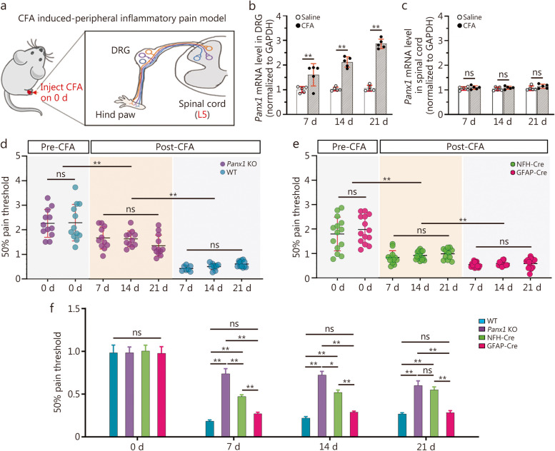
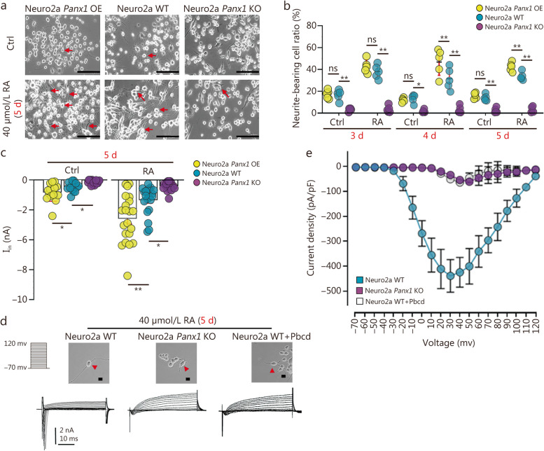
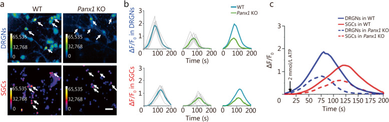
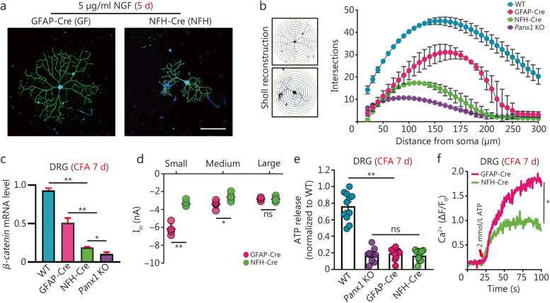
Methods: Wild-type (WT, n = 24), global Panx1 KO (n = 24), neuron-specific Panx1 KO (n = 20), and glia-specific Panx1 KO (n = 20) mice were used in this study at Albert Einstein College of Medicine. The von Frey test was used to quantify pain sensitivity in these mice following complete Freund's adjuvant (CFA) injection (7, 14, and 21 d). The qRT-PCR was employed to measure mRNA levels of Panx1, Panx2, Panx3, Cx43, Calhm1, and β-catenin. Laser scanning confocal microscopy imaging, Sholl analysis, and electrophysiology were utilized to evaluate the impact of Panx1 on neuronal excitability and morphology in Neuro2a and dorsal root ganglion neurons (DRGNs) in which Panx1 expression or function was manipulated. Ethidium bromide (EtBr) dye uptake assay and calcium imaging were employed to investigate the role of Panx1 in adenosine triphosphate (ATP) sensitivity. β-galactosidase (β-gal) staining was applied to determine the relative cellular expression levels of Panx1 in trigeminal ganglia (TG) and DRG of transgenic mice.

Results: Global or neuron-specific Panx1 deletion markedly decreased pain thresholds after CFA stimuli (7, 14, and 21 d; P < 0.01 vs. WT group), indicating that Panx1 was positively correlated with pain sensitivity. In Neuro2a, global Panx1 deletion dramatically reduced neurite extension and inward currents compared to the WT group (P < 0.05), revealing that Panx1 enhanced neurogenesis and excitability. Similarly, global Panx1 deletion significantly suppressed Wnt/β-catenin dependent DRG neurogenesis following 5 d of nerve growth factor (NGF) treatment (P < 0.01 vs. WT group). Moreover, Panx1 channels enhanced DRG neuron response to ATP after CFA injection (P < 0.01 vs. Panx1 KO group). Furthermore, ATP release increased Ca2+ responses in DRGNs and satellite glial cells surrounding them following 7 d of CFA treatment (P < 0.01 vs. Panx1 KO group), suggesting that Panx1 in glia also impacts exaggerated neuronal excitability. Interestingly, neuron-specific Panx1 deletion was found to markedly reduce differentiation in cultured DRGNs, as evidenced by stunted neurite outgrowth (P < 0.05 vs. Panx1 KO group; P < 0.01 vs. WT group or GFAP-Cre group), blunted activation of Wnt/β-catenin signaling (P < 0.01 vs. WT, Panx1 KO and GFAP-Cre groups), and diminished cell excitability (P < 0.01 vs. GFAP-Cre group) and response to ATP stimulation (P < 0.01 vs. WT group). Analysis of β-gal staining showed that cellular expression levels of Panx1 in neurons are significantly higher (2.5-fold increase) in the DRG than in the TG.

Conclusions: The present study revealed that neuronal Panx1 is a prominent driver of peripheral sensitivity in the setting of inflammatory pain through cell-autonomous effects on neuronal excitability. This hyperexcitability dependence on neuronal Panx1 contrasts with inflammatory orofacial pain, where similar studies revealed a prominent role for glial Panx1. The apparent differences in Panx1 expression in neuronal and non-neuronal TG and DRG cells are likely responsible for the distinct impact of these cell types in the two pain models.

Keywords: Dorsal root ganglion; Panx1; Peripheral sensitization; Plantar inflammatory pain; Satellite glial cell.


Conflict of interest statement

The authors declare that they have no competing interests.

Figures

Fig. 1
Fig. 1
Global or neuronal Panx1 deletion reduces allodynia while GFAP-targeted deletion does not. a Schematic plot of CFA induced peripheral inflammatory pain model. Panx1 mRNA expression in DRG (b) and spinal cord (c) from WT and Panx1 KO mice within 3 weeks after CFA inflammation. d Three-week dynamic 50% pain threshold of WT and Panx1 KO mice. e Three-week dynamic 50% pain threshold of NFH-Cre and GFAP-Cre Panx1 KO mice. f Comparison of pain sensitivity changes among those four genotypes above at each time point. For panels b and c, n = 3 in saline and CFA group; n = 11 in WT group, n = 11 in Panx1 KO group, n = 12 in GFAP-Cre group, and n = 12 in NFH-Cre group. Two-way ANOVA with Bonferroni, interaction F (2, 12) = 26.07 in panel b, F (2, 12) = 0.1176 in panel c, F (7, 68) = 29.66 in panel d, F (7, 87) = 33.44 in panel e, and F (9, 170) = 5.641 in panel f. ns non-significant, *P < 0.05, **P < 0.01. CFA complete Freund’s adjuvant, DRG dorsal root ganglion, NFH-Cre neuro filament H-Cre, GFAP-Cre glial fibrillary acidic protein-Cre, Panx1 pannexin 1, Panx1 KO pannexin 1 knockout, WT wild-type
Fig. 2
Fig. 2
Panx1 facilitates neurite extension and promotes cell excitability in Neuro2a neuroblastoma cells. a Representative photomicrograph of Neuro2a WT, Neuro2a Panx1 KO, and Neuro2a Panx1 OE cells after 5 d of RA treatment. Arrows indicate processes of representative cells in each condition. b Neurite extension analysis in WT, Panx1-deleted, and Neuro2a Panx1 OE cells at d 3–5 in culture with or without 40 µmol/L RA (n = 5). c Quantification of peak inward currents in Neuro2a WT, Neuro2a Panx1 KO and Neuro2a Panx1 OE on day 5 with and without RA treatment (n = 15–22 cells in each group). d Representative recordings of whole-cell voltage-clamp currents in Neuro2a WT, Neuro2a Panx1 KO, and Neuro2a WT plus Panx1 blocker 1 mmol/L Pbcd following 5 d treatment with RA. e Current density analysis of Neuro2a cells with 1 mmol/L Pbcd (n = 9–11 in each group). Two-way ANOVA with Bonferroni in b, c, interaction F (2, 24) = 15.04 in panel b, F (2, 56) = 18.62 in panel c; scale bar = 150 μm in panel a and scale bar = 20 μm in panel d. ns non-significant, *P < 0.05, **P < 0.01. Neuro2a WT neuro2a wild-type, Neuro2a Panx1 KO neuro2a pannexin 1 knockout, Neuro2a Panx1 OE pannexin 1-overexpressing neuro2a, Pbcd probenecid, RA retinoic acid
Fig. 3
Fig. 3
Global Panx1 deletion impacts DRG neurogenesis via Wnt/β-catenin pathway. a cell morphology of NF-200 stained DRG neuron from WT and Panx1 KO mice after 5 d of NGF treatment. b Sholl analysis of intersection numbers in WT and Panx1 KO neurons (WT vs. Panx1 KO group, P < 0.01). c Following 5 d of treatment with NGF, cell morphology of DRG neurons (DRGNs) from WT and Panx1 KO mice was visualized NF200. Exposure to 20 mmol/L LiCl, 30 nmol/L IWP2, and 100 nmol/L Triptonide was compared. d mRNA expression of β-catenin in WT and Panx1 KO DRGNs after 5 d of treatment with NGF. Exposure to LiCl, IWP2, and Triptonide was also compared. e Quantification of cell morphology indicated neurite lengths in WT and Panx1 KO DRGNs following treatment with modifiers of the Wnt/β-catenin pathway (n = 15 in each group). Two-way ANOVA with Bonferroni in d, e, interaction F (7, 40) = 2.595 in panel d, F (3, 16) = 183.1 in panel e. ns non-significant, *P < 0.05, **P < 0.01. Scale bar = 100 μm in panels a, and scale bar = 20 μm in panels c. DRG dorsal root ganglion, IWP2 Wnt production inhibitor 2, LiCl lithium chloride, NF200 neurofilament 200, NGF nerve growth factor, WT wild-type, Panx1 KO pannexin 1 knockout
Fig. 4
Fig. 4
Panx1 enhances NGF-induced inward currents, dye uptake, and ATP release. a After 5 d of NGF treatment, representative electrophysiological recordings of different diameter-DRGNs [small (< 20 μm), medium (20–25 μm), and large (> 25 μm)] from WT and Panx1 KO mice were detected and analyzed (b). c Maximum inward currents recordings from WT and Panx1 KO sensory neurons before and 5 d after NGF treatment (Ctrl, 5% serum treated; 5 µg/ml NGF treated; n = 10). d Representative recordings from WT, Panx1 KO and Pbcd inhibited Panx1 groups (n = 10). e Current density analysis of those three types of DRGNs above. e Statistical analysis of c. f EtBr dye uptake images and data analysis (g) between WT and Panx1 KO NGF treated DRGNs with and without 2 mmol/L ATP treatment. h ATP release levels analysis between WT and Panx1 KO DRGNs following CFA injection on day 7. Scale bar = 20 μm in d, f. ns non-significant, *P < 0.05, **P < 0.01 for one-way ANOVA in b, c, g, h. Ctrl control, DRGNs dorsal root ganglion neurons, EtBr ethidium bromide, NGF nerve growth factor, WT wild-type, Panx1 KO pannexin 1 knockout, Pbcd probenecid
Fig. 5
Fig. 5
Both dorsal root ganglion neurons (DRGNs) and satellite glial cells (SGCs) show heightened sensitivity to ATP in co-cultures from CFA-treated mice. a Ca2+ imaging of Fura2-loaded dissociated WT and Panx1 KO DRGNs and SGCs. Scale bar = 50 μm. b Quantification of calcium responses from DRGNs (blue) and SGCs (red) [n = 5; statistical analysis from either DRGNs or SGCs indicates that there are stronger calcium waves in WT, compared to Panx1 KO group (P < 0.01)]. c Data summarized and replotted from data sets in b (downward-pointing arrow indicates a time of ATP application). ns non-significant, *P < 0.05, **P < 0.01 for one-way ANOVA with repeated measures in c. WT wild-type, Panx1 KO pannexin 1 knockout
Fig. 6
Fig. 6
Panx1 contributes to DRG changes in both neurons and glia in chronic inflammatory pain. a Representative morphologies of dorsal root ganglion neurons (DRGNs) from GFAP-Cre and NFH-Cre mice stained with NF200 antibody (green) after 5 d of NGF induction. Scale bar = 100 μm. b Sholl analysis of NGF-treated GFAP-Cre and NFH-Cre DRGNs on day 5. cβ-catenin mRNA analysis of those NGF-treated GFAP-Cre and NFH-Cre DRGNs compared to WT and global Panx1 KO [n = 6; NFH-Cre vs. GFAP-Cre group (P < 0.01), global Panx1 KO vs. NFH-Cre group (P < 0.05), global Panx1 KO vs. GFAP-Cre group (P < 0.01), WT vs. NFH-Cre or GFAP-Cre or global Panx1 KO group (P < 0.01)]. d Quantification of inward currents from GFAP-Cre and NFH-Cre DRGNs (traced with DiI) projecting to CFA-injected hind paw on day 7 (n = 8). e ATP release analysis of WT, Panx1 KO, GFAP-Cre and NFH-Cre DRG ganglia 7 d post-CFA injection (n = 10). f Ca2+ response analysis of GFAP-Cre and NFH-Cre DRGNs after CFA injection on day 7 (n = 5). ns non-significant, *P < 0.05, **P < 0.01 for one-way ANOVA in b, c, d, e, f with repeated measures. CFA complete Freund’s adjuvant, Panx1 pannexin 1, Panx1 KO pannexin 1 knockout, DRG dorsal root ganglion, GFAP-Cre glial fibrillary acidic protein-Cre, NFH-Cre neuro filament H-Cre, NF200 neurofilament 200, NGF nerve growth factor, WT wild-type

Comment in

References

    1. Meacham K, Shepherd A, Mohapatra DP, Haroutounian S. Neuropathic pain: central vs. peripheral mechanisms. Curr Pain Headache Rep. 2017;21(6):28. doi: 10.1007/s11916-017-0629-5. - DOI - PubMed
    1. Ji RR, Nackley A, Huh Y, Terrando N, Maixner W. Neuroinflammation and central sensitization in chronic and widespread pain. Anesthesiology. 2018;129(2):343–66. doi: 10.1097/ALN.0000000000002130. - DOI - PMC - PubMed
    1. Gold MS, Gebhart GF. Nociceptor sensitization in pain pathogenesis. Nat Med. 2010;16(11):1248–57. doi: 10.1038/nm.2235. - DOI - PMC - PubMed
    1. Enthoven WTM, Roelofs PD, Koes BW. NSAIDs for chronic low back pain. JAMA. 2017;317(22):2327–8. doi: 10.1001/jama.2017.4571. - DOI - PubMed
    1. Morrone AL, Scuteri D, Rombola L, Mizoguchi H, Bagetta G. Opioids resistance in chronic pain management. Curr Neuropharmacol. 2017;15(3):444–56. doi: 10.2174/1570159X14666161101092822. - DOI - PMC - PubMed

Publication types