Multicenter Study
. 2024 Sep;120 Suppl 1(Suppl 1):S41-S50.
doi: 10.1016/j.ajcnut.2024.04.027. Epub 2024 Apr 27.

Duodenal quantitative mucosal morphometry in children with environmental enteric dysfunction: a cross-sectional multicountry analysis

Collaborators, Affiliations
Multicenter Study

Duodenal quantitative mucosal morphometry in children with environmental enteric dysfunction: a cross-sectional multicountry analysis

Lubaina Ehsan et al. Am J Clin Nutr. 2024 Sep.

Abstract

Background: Environmental enteric dysfunction (EED), a chronic inflammatory condition of the small intestine, is an important driver of childhood malnutrition globally. Quantifying intestinal morphology in EED allows for exploration of its association with functional and disease outcomes.

Objectives: We sought to define morphometric characteristics of childhood EED and determine whether morphology features were associated with disease pathophysiology.

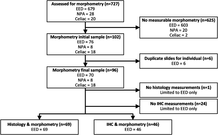
Methods: Morphometric measurements and histology were assessed on duodenal biopsy slides for this cross-sectional study from children with EED in Bangladesh, Pakistan, and Zambia (n = 69), and those with no pathologic abnormality (NPA; n = 8) or celiac disease (n = 18) in North America. Immunohistochemistry was also conducted on 46, 8, and 18 biopsy slides, respectively. Linear mixed-effects regression models were used to reveal morphometric differences between EED compared with NPA or celiac disease and identify associations between morphometry and histology or immunohistochemistry among children with EED.

Results: In duodenal biopsies, median EED villus height (248 μm), crypt depth (299 μm), and villus:crypt (V:C) ratio (0.9) values ranged between those of NPA (396 μm villus height; 246 μm crypt depth; 1.6 V:C ratio) and celiac disease (208 μm villus height; 365 μm crypt depth; 0.5 V:C ratio). Among EED biopsy slides, morphometric assessments were not associated with histologic parameters or immunohistochemical markers, other than pathologist-determined subjective semiquantitative villus architecture.

Conclusions: Morphometric analysis of duodenal biopsy slides across geographies identified morphologic features of EED, specifically short villi, elongated crypts, and a smaller V:C ratio relative to NPA slides, although not as severe as in celiac slides. Morphometry did not explain other EED features, suggesting that EED histopathologic processes may be operating independently of morphology. Although acknowledging the challenges with obtaining relevant tissue, these data form the basis for further assessments of the role of morphometry in EED.

Keywords: crypt; gastrointestinal morphology; global health; pediatric; villi.


Figures

FIGURE 1
FIGURE 1
Villus length and crypt depth morphometry measurements from duodenal biopsy images. (A) Illustration of villus and crypt structures that were identified for morphometric measurements. (B) Mucosal morphometry was measured from villus–crypt units present in individual-derived duodenal biopsy tissue. Top image: Villus length was measured from the tip of the villi to the “shoulder” of the villus in the crypt–villus junction, marking the start of the crypt (height). Crypt depth was measured from the “shoulder” of the crypt–villus junction to the base of the crypt (depth). Arrow: the region of interest (ROI) for visualizing the villus–crypt junction, or the end of the mucosa or beginning of the submucosa, which enables the assessment of the depth of the mucosa. Bottom image: Morphometric measurement where crypt depth was measured taking the curvature of the crypt into account.
FIGURE 2
FIGURE 2
Flowchart depiction of the biopsy slides obtained and used within this study. A total of 727 duodenal biopsy slides were obtained from children enrolled at 4 EEDBI Consortium centers (BEECH, Biomarkers of Environmental Enteropathy in Children, University Teaching Hospital, Zambia; BEED, Bangladesh Environmental Enteric Dysfunction, International Centre for Diarrhoeal Disease Research, Bangladesh; SEEM, Study of Environmental Enteropathy and Malnutrition, Aga Khan University, Pakistan; CCHMC, Cincinnati Children's Hospital Medical Center, Cincinnati, United States). Slides where no morphometric measurements were obtained and duplicate slides per individual were excluded from our analysis. Environmental enteric dysfunction (EED) slides were further analyzed for histology, morphometry, and immunohistochemistry.
FIGURE 3
FIGURE 3
Morphometric measurement examples from across sites. Top row right to left: SEEM, BEED, and BEECH. Bottom row right to left: CCHMC Celiac and CCHMC NPA. Measurements within red boxes indicate crypt depth whereas those within yellow boxes indicate villus height. Region of interest (ROI, as described in methods) has been drawn in blue for visualizing the villus–crypt junction. Complete crypt depth was not evident for SEEM, BEED, BEECH, and NPA as shown in the images due to which measurements were extended to the muscularis mucosa as complete crypt depth was not evident to reduce the risk of crypt underestimation, as detailed in the methods section. Abbreviations: BEECH, Biomarkers of Environmental Enteropathy in Children, University Teaching Hospital, Zambia; BEED, Bangladesh Environmental Enteric Dysfunction, International Centre for Diarrhoeal Disease Research, Bangladesh (icddr,b); CCHMC, Cincinnati Children's Hospital Medical Center, Cincinnati, United States; NPA, no pathologic abnormality; SEEM, Study of Environmental Enteropathy and Malnutrition, Aga Khan University, Pakistan.

Similar articles

Cited by

References

    1. Denno D.M., Tarr P.I., Nataro J.P. Environmental enteric dysfunction: a case definition for intervention trials. Am. J. Trop. Med. Hyg. 2017;97(6):1643–1646. doi: 10.4269/ajtmh.17-0183. - DOI - PMC - PubMed
    1. Kosek M.N. MAL-ED Network Investigators, Causal pathways from enteropathogens to environmental enteropathy: findings from the MAL-ED birth cohort study. EBioMedicine. 2017;18:109–117. doi: 10.1016/j.ebiom.2017.02.024. - DOI - PMC - PubMed
    1. Humphrey J.H. Child undernutrition, tropical enteropathy, toilets, and handwashing. Lancet. 2009;374(9694):1032–1035. doi: 10.1016/S0140-6736(09)60950-8. - DOI - PubMed
    1. Jiang N.M., Tofail F., Moonah S.N., Scharf R.J., Taniuchi M., Ma J.Z., et al. Febrile illness and pro-inflammatory cytokines are associated with lower neurodevelopmental scores in Bangladeshi infants living in poverty. BMC Pediatr. 2014;14:50. https://doi.org/1186/1471-2431-14-50. - PMC - PubMed
    1. Prendergast A.J., Humphrey J.H. The stunting syndrome in developing countries. Paediatr. Int. Child. Health. 2014;34(4):250–265. doi: 10.1179/2046905514Y.0000000158. - DOI - PMC - PubMed

Publication types

LinkOut - more resources