

Super-selective Embolisation and Surgical Excision of the Facial Arteriovenous Malformation
- PMID: 38686263
- PMCID: PMC11056801
- DOI: 10.7759/cureus.57240
Super-selective Embolisation and Surgical Excision of the Facial Arteriovenous Malformation
Abstract
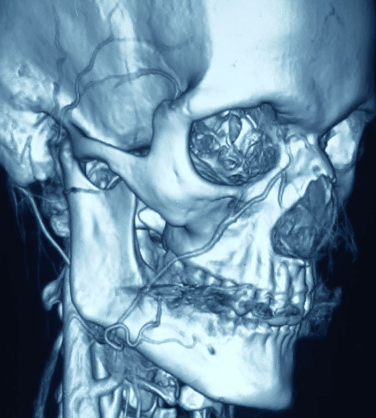
Vascular anomalies broadly include vascular tumours and malformations. Arteriovenous malformations (AVM), though rare in the oral and maxillofacial regions, can present with swelling, facial asymmetry, ulceration, and bleeding tendencies, which can be life-threatening. Thus, to minimise the associated life-threatening consequences, prompt and appropriate diagnosis of the lesion is necessitated. The management of the AVM is a therapeutic challenge for maxillofacial surgeons; however, technological advances in interventional radiology have gained a foothold. Super-selective embolisation of the feeder vessels with subsequent resection of the lesion is the most widely accepted approach for management. The present report describes a unique case of a facial AVM managed through a trans-oral approach without any post-operative sequelae.
Keywords: arterivenous malformation; face; intraoral approach; resection; superselective embolisation.
Copyright © 2024, Chaulagain et al.
Conflict of interest statement
The authors have declared that no competing interests exist.
Figures


References
-
- Hemangiomas and vascular malformations in infants and children: a classification based on endothelial characteristics. Mulliken JB, Glowacki J. Plast Reconstr Surg. 1982;69:412–422. - PubMed
-
- Vascular malformations: classification, diagnosis and treatment. Carqueja IM, Sousa J, Mansilha A. Int Angiol. 2018;37:127–142. - PubMed
-
- Current management of hemangiomas and vascular malformations. Marler JJ, Mulliken JB. Clin Plast Surg. 2005;32:99-116, ix. - PubMed
-
- Hemangiomas, vascular malformations, and lymphovenous malformations: classification and methods of treatment. Jackson IT, Carreño R, Potparic Z, Hussain K. Plast Reconstr Surg. 1993;91:1216–1230. - PubMed
Publication types
LinkOut - more resources
Full Text Sources