Case Reports
. 2024 Apr 15:15:1243566.
doi: 10.3389/fimmu.2024.1243566. eCollection 2024.

Lichen planus pemphigoides with predominant mucous membrane involvement: a series of 12 patients and a literature review

Affiliations
Case Reports

Lichen planus pemphigoides with predominant mucous membrane involvement: a series of 12 patients and a literature review

Loraine Combemale et al. Front Immunol. .

Abstract

Background: Lichen planus pemphigoides (LPP), an association between lichen planus and bullous pemphigoid lesions, is a rare subepithelial autoimmune bullous disease. Mucous membrane involvement has been reported previously; however, it has never been specifically studied.

Methods: We report on 12 cases of LPP with predominant or exclusive mucous membrane involvement. The diagnosis of LPP was based on the presence of lichenoid infiltrates in histology and immune deposits in the basement membrane zone in direct immunofluorescence and/or immunoelectron microscopy. Our systematic review of the literature, performed according to the Preferred Reporting Items for Systematic Reviews and Meta-Analyses guidelines, highlights the clinical and immunological characteristics of LPP, with or without mucous membrane involvement.

Results: Corticosteroids are the most frequently used treatment, with better outcomes in LPP with skin involvement alone than in that with mucous membrane involvement. Our results suggest that immunomodulators represent an alternative first-line treatment for patients with predominant mucous membrane involvement.

Keywords: autoimmune blistering dermatosis; autoimmune blistering disease; bullous pemphigoid; lichen planus pemphigoides; mucous membrane pemphigoid; oral lichen planus.


Conflict of interest statement

The authors declare that the research was conducted in the absence of any commercial or financial relationships that could be construed as a potential conflict of interest.

Figures
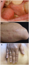
Figure 1
Figure 1
Typical clinical lichenoid lesions. (A) White reticulated network on buccal mucosa (patient N°9), (B) cutaneous violaceous papules with Wickham network on a left forearm (patient N°2), and (C) lichen planus onychodystrophy (pterygium) of one patient (patient N°12).
Figure 2
Figure 2
Typical clinical bullous lesions. (A) Blisters and white reticulated network observed on buccal mucosa (patient N°3) and (B) bullous and erythematous lesions on a leg (patient N°4).
Figure 3
Figure 3
Erythema (black arrows), erosion, white lichenoid lesion, and atrophy on vestibular gingiva. Erythema could be a manifestation of lichen planus or mucous membrane pemphigoid. (patient N°12).
Figure 4
Figure 4
Histology of mucous membrane biopsy (Hematoxylin and eosin staining) and subepithelial cleavage and lichenoid reaction pattern (band-like lymphocytic infiltrate hugging the chorioepithelial junction with, depending on the patient, vacuolar degeneration of the basal layer of the epithelium, necrosis of individual keratinocytes, and lymphocytic exocytosis). (A, B) Both in one section of a biopsy (original magnification, 50×), with (A) discrete subepithelial cleavage and predominant lichenoid reaction (patient N° 8) and (B) large subepithelial cleavage and discrete lichenoid pattern with lymphocytic exocytosis (patient N° 12). (C, D) Separately in two different sections of one biopsy (original magnification, 100×), with (C) subepithelial cleavage and (D) a lichenoid reaction pattern (patient N°3). (E, F) Separately in two different biopsies (original magnification, 100×), with (E) subepithelial cleavage on gingiva biopsy and (F) a lichenoid reaction pattern on buccal mucosa biopsy (patient N°5). (G, H) Direct immunofluorescence, with (G) linear immune deposits of IgG on the DEJ (patient N°11) and (H) on the CEJ (patient N°4).
Figure 5
Figure 5
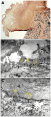
Direct immunoelectron microscopy. (A) Semi-thin section demonstrating linear deposits of autoantibodies on the chorioepithelial junction (original magnification, 40×; patient N°2). (B) Ultra-thin section demonstrating autoimmune deposits on hemidesmosomes or hemidesmosomes and lamina lucida (shown by the black arrows) (original magnification, 15,000×; patient N°4). (C) Ultra-thin section demonstrating thick immune deposits (shown by the black arrows) on lamina densa intermittently overlying the lamina lucida (original magnification, 15,000×; patient N°1).

References

    1. Kaposi M. Lichen ruber pemphigoides. Arch J Dermatol Syph. (1892) 24:343–6.
    1. Stingl G, Hobular K. Coexistence of lichen planus and bullous pemphigoid. A immunopathological study. Br J Dermatol. (1975) 93:313–20. doi: 10.1111/j.1365-2133.1975.tb06497.x - DOI - PubMed
    1. Cognat T, Gayrard L, Adam C, Balmex B, MaChado P, Nicolas JF, et al. . Lichen plan pemphigoïde [Lichen planus pemphigoides]. Ann Dermatol Venereol. (1991) 118:387–90. - PubMed
    1. Ogg GS, Bhogal BS, Hashimoto T, Coleman R, Barker JN. Ramipril-associated lichen planus pemphigoides. Br J Dermatol. (1997) 136:412–4. - PubMed
    1. Zillikens D, Caux F, Mascaro JM, Wesselmann U, Schmidt E, Prost C, et al. . Autoantibodies in lichen planus pemphigoides react with a novel epitope within the C-terminal NC16A domain of BP180. J Invest Dermatol. (1999) 113:117–21. doi: 10.1046/j.1523-1747.1999.00618.x - DOI - PubMed

Publication types

LinkOut - more resources