Single-cell transcriptomic profiling of the neonatal oviduct and uterus reveals new insights into upper Müllerian duct regionalization
- PMID: 38686936
- PMCID: PMC11095678
- DOI: 10.1096/fj.202400303R
Single-cell transcriptomic profiling of the neonatal oviduct and uterus reveals new insights into upper Müllerian duct regionalization
Abstract
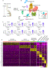
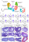
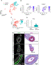
The upper Müllerian duct (MD) is patterned and specified into two morphologically and functionally distinct organs, the oviduct and uterus. It is known that this regionalization process is instructed by inductive signals from the adjacent mesenchyme. However, the interaction landscape between epithelium and mesenchyme during upper MD development remains largely unknown. Here, we performed single-cell transcriptomic profiling of mouse neonatal oviducts and uteri at the initiation of MD epithelial differentiation (postnatal day 3). We identified major cell types including epithelium, mesenchyme, pericytes, mesothelium, endothelium, and immune cells in both organs with established markers. Moreover, we uncovered region-specific epithelial and mesenchymal subpopulations and then deduced region-specific ligand-receptor pairs mediating mesenchymal-epithelial interactions along the craniocaudal axis. Unexpectedly, we discovered a mesenchymal subpopulation marked by neurofilaments with specific localizations at the mesometrial pole of both the neonatal oviduct and uterus. Lastly, we analyzed and revealed organ-specific signature genes of pericytes and mesothelial cells. Taken together, our study enriches our knowledge of upper MD development, and provides a manageable list of potential genes, pathways, and region-specific cell subtypes for future functional studies.
Keywords: Müllerian duct; craniocaudal patterning; oviduct; regionalization; uterus.
© 2024 The Authors. The FASEB Journal published by Wiley Periodicals LLC on behalf of Federation of American Societies for Experimental Biology.
Figures


Update of
-
Single-cell transcriptomic profiling of the neonatal oviduct and uterus reveals new insights into upper Müllerian duct regionalization.bioRxiv [Preprint]. 2023 Dec 23:2023.12.20.572607. doi: 10.1101/2023.12.20.572607. bioRxiv. 2023. Update in: FASEB J. 2024 May 15;38(9):e23632. doi: 10.1096/fj.202400303R. PMID: 38187777 Free PMC article. Updated. Preprint.
Similar articles
-
Decoding Müllerian Duct Epithelial Regionalization.Mol Reprod Dev. 2025 Feb;92(2):e70018. doi: 10.1002/mrd.70018. Mol Reprod Dev. 2025. PMID: 39994938 Free PMC article. Review.
-
Single-cell transcriptomic profiling of the neonatal oviduct and uterus reveals new insights into upper Müllerian duct regionalization.bioRxiv [Preprint]. 2023 Dec 23:2023.12.20.572607. doi: 10.1101/2023.12.20.572607. bioRxiv. 2023. Update in: FASEB J. 2024 May 15;38(9):e23632. doi: 10.1096/fj.202400303R. PMID: 38187777 Free PMC article. Updated. Preprint.
-
Retinoic acid signaling determines the fate of uterine stroma in the mouse Müllerian duct.Proc Natl Acad Sci U S A. 2016 Dec 13;113(50):14354-14359. doi: 10.1073/pnas.1608808113. Epub 2016 Nov 22. Proc Natl Acad Sci U S A. 2016. PMID: 27911779 Free PMC article.
-
CTNNB1 in mesenchyme regulates epithelial cell differentiation during Müllerian duct and postnatal uterine development.Mol Endocrinol. 2013 Sep;27(9):1442-54. doi: 10.1210/me.2012-1126. Epub 2013 Jul 31. Mol Endocrinol. 2013. PMID: 23904126 Free PMC article.
-
Normal and abnormal epithelial differentiation in the female reproductive tract.Differentiation. 2011 Oct;82(3):117-26. doi: 10.1016/j.diff.2011.04.008. Epub 2011 May 25. Differentiation. 2011. PMID: 21612855 Free PMC article. Review.
Cited by
-
Uterine organoids reveal insights into epithelial specification and plasticity in development and disease.Proc Natl Acad Sci U S A. 2025 Feb 4;122(5):e2422694122. doi: 10.1073/pnas.2422694122. Epub 2025 Jan 30. Proc Natl Acad Sci U S A. 2025. PMID: 39883834 Free PMC article.
-
Decoding Müllerian Duct Epithelial Regionalization.Mol Reprod Dev. 2025 Feb;92(2):e70018. doi: 10.1002/mrd.70018. Mol Reprod Dev. 2025. PMID: 39994938 Free PMC article. Review.
References
-
- Stewart CA, and Behringer RR (2012) Mouse oviduct development. Results Probl Cell Differ 55, 247–262 - PubMed
-
- Robboy SJ, Szyfelbein WM, Goellner JR, Kaufman RH, Taft PD, Richard RM, Gaffey TA, Prat J, Virata R, Hatab PA, McGorray SP, Noller KL, Townsend D, Labarthe D, and Barnes AB (1981) Dysplasia and cytologic findings in 4,589 young women enrolled in diethylstilbestrol-adenosis (DESAD) project. Am J Obstet Gynecol 140, 579–586 - PubMed
Publication types
MeSH terms
Grants and funding
LinkOut - more resources
Full Text Sources
Molecular Biology Databases

