. 2024 Apr;311(1):e231991.
doi: 10.1148/radiol.231991.

Addition of Contrast-enhanced Mammography to Tomosynthesis for Breast Cancer Detection in Women with a Personal History of Breast Cancer: Prospective TOCEM Trial Interim Analysis

Affiliations

Addition of Contrast-enhanced Mammography to Tomosynthesis for Breast Cancer Detection in Women with a Personal History of Breast Cancer: Prospective TOCEM Trial Interim Analysis

Wendie A Berg et al. Radiology. 2024 Apr.

Abstract

Background Digital breast tomosynthesis (DBT) is often inadequate for screening women with a personal history of breast cancer (PHBC). The ongoing prospective Tomosynthesis or Contrast-Enhanced Mammography, or TOCEM, trial includes three annual screenings with both DBT and contrast-enhanced mammography (CEM). Purpose To perform interim assessment of cancer yield, stage, and recall rate when CEM is added to DBT in women with PHBC. Materials and Methods From October 2019 to December 2022, two radiologists interpreted both examinations: Observer 1 reviewed DBT first and then CEM, and observer 2 reviewed CEM first and then DBT. Effects of adding CEM to DBT on incremental cancer detection rate (ICDR), cancer type and node status, recall rate, and other performance characteristics of the primary radiologist decisions were assessed. Results Among the participants (mean age at entry, 63.6 years ± 9.6 [SD]), 1273, 819, and 227 women with PHBC completed year 1, 2, and 3 screening, respectively. For observer 1, year 1 cancer yield was 20 of 1273 (15.7 per 1000 screenings) for DBT and 29 of 1273 (22.8 per 1000 screenings; ICDR, 7.1 per 1000 screenings [95% CI: 3.2, 13.4]) for DBT plus CEM (P < .001). Year 2 plus 3 cancer yield was four of 1046 (3.8 per 1000 screenings) for DBT and eight of 1046 (7.6 per 1000 screenings; ICDR, 3.8 per 1000 screenings [95% CI: 1.0, 7.6]) for DBT plus CEM (P = .001). Year 1 recall rate for observer 1 was 103 of 1273 (8.1%) for (incidence) DBT alone and 187 of 1273 (14.7%) for DBT plus CEM (difference = 84 of 1273, 6.6% [95% CI: 5.3, 8.1]; P < .001). Year 2 plus 3 recall rate was 40 of 1046 (3.8%) for DBT and 92 of 1046 (8.8%) for DBT plus CEM (difference = 52 of 1046, 5.0% [95% CI: 3.7, 6.3]; P < .001). In 18 breasts with cancer detected only at CEM after integration of both observers, 13 (72%) cancers were invasive (median tumor size, 0.6 cm) and eight of nine (88%) with staging were N0. Among 1883 screenings with adequate reference standard, there were three interval cancers (one at the scar, two in axillae). Conclusion CEM added to DBT increased early breast cancer detection each year in women with PHBC, with an accompanying approximately 5.0%-6.6% recall rate increase. Clinical trial registration no. NCT04085510 © RSNA, 2024 Supplemental material is available for this article.


Conflict of interest statement

Disclosures of conflicts of interest: W.A.B. Institution received grants from Koios Medical and Pennsylvania Breast Cancer Coalition; author received consulting fees from Exai Bio; voluntary chief scientific advisor, DenseBreast-info.org; voluntary associate editor, Journal of Breast Imaging. J.M.B. No relevant relationships. A.I.B. Institution received grants from NIH, Pennsylvania Breast Cancer Coalition, DOD, BCRF, author received consulting fees from Hologic. A.V. No relevant relationships. D.M.C. No relevant relationships. A.H.L. Consulting fees for Hologic review study. M.A.G. No relevant relationships. A.E.K. No relevant relationships. B.E.N. No relevant relationships. J.Y.H. No relevant relationships. U.W. No relevant relationships. C.M.H. No relevant relationships. K.S.H. No relevant relationships. R.F.R. No relevant relationships. D.D.S. No relevant relationships. B.A.C. No relevant relationships. C.S.C. No relevant relationships. L.P.W. No relevant relationships. J.H.S. No relevant relationships. M.L.Z. NIH/NCI R01 CA258898-01A1 PI NIH/NCI R01 CA237827 MPI Hologic, PI of institutional grant Bayer, PI of institutional grant; Applied Radiology honoraria for forum participation; FDA Radiological Device Panel Member Panel meeting 11/7/2023; NAPBC board member.

Figures

None
Graphical abstract
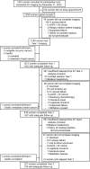
Flowchart of study participants. Adequate follow-up was biopsy
diagnosis of cancer or no breast cancer diagnosis at surgery or follow-up
imaging of at least 10.5 months, or, if no subsequent imaging or surgery had
been performed, clinical follow-up of at least 12 months. Four women had
mild contrast agent reactions in year 2 and planned to premedicate and
continue participation, but one failed to premedicate and was excluded in
year 3. eGFR = estimated glomerular filtration rate, IV =
intravenous.
Figure 1:
Flowchart of study participants. Adequate follow-up was biopsy diagnosis of cancer or no breast cancer diagnosis at surgery or follow-up imaging of at least 10.5 months, or, if no subsequent imaging or surgery had been performed, clinical follow-up of at least 12 months. Four women had mild contrast agent reactions in year 2 and planned to premedicate and continue participation, but one failed to premedicate and was excluded in year 3. eGFR = estimated glomerular filtration rate, IV = intravenous.
Images in a 67-year-old woman with triple receptor–negative
invasive ductal carcinoma (IDC) seen only at contrast-enhanced mammography
(CEM) at year 2. (A) Left craniocaudal (CC) (left) and mediolateral oblique
(MLO) (right) low-energy images show scattered fibroglandular density and
postsurgical scarring, with clips in the lower inner quadrant at the site of
lumpectomy for a 2.1-cm grade 3 IDC, estrogen receptor– and
progesterone receptor–positive and human epidermal growth factor
receptor 2 (HER2) (ERBB2 gene)–negative lesion 11 years prior.
Scattered benign-appearing calcifications are noted. The participant also
completed radiation therapy and adjuvant chemotherapy and was treated with
tamoxifen for 7 years and then with an aromatase inhibitor for 3 years, with
last use 1 year prior to study entry. (B) Recombined CC (left) and MLO
(right) CEM images obtained in year 2 show moderately conspicuous
enhancement of an oval mass in the upper outer left breast (arrows), which
was new from the prior CEM examination (not shown). This lesion was assessed
as Breast Imaging Reporting and Data System (BI-RADS) 4B, moderately
suspicious, by observer 1 and as BI-RADS 3, probably benign, but recommended
for additional evaluation, by observer 2. At the time, CEM-guided biopsy was
not available, so the participant underwent MRI and MRI-guided biopsy. (C)
Axial maximum intensity projection from T1-weighted fat-suppressed MRI
(left) shows moderately intense enhancement of the same mass (arrow), with
plateau and washout kinetics (arrow) on axial post-contrast fat-suppressed
T1-weighted image with kinetic overlay (right). MRI-guided biopsy and
excision revealed a 0.5-cm grade 3 IDC, triple receptor–negative
lesion (Ki-67 proliferation index of 55%). Three sentinel nodes were
negative for metastasis.
Figure 2:
Images in a 67-year-old woman with triple receptor–negative invasive ductal carcinoma (IDC) seen only at contrast-enhanced mammography (CEM) at year 2. (A) Left craniocaudal (CC) (left) and mediolateral oblique (MLO) (right) low-energy images show scattered fibroglandular density and postsurgical scarring, with clips in the lower inner quadrant at the site of lumpectomy for a 2.1-cm grade 3 IDC, estrogen receptor– and progesterone receptor–positive and human epidermal growth factor receptor 2 (HER2) (ERBB2 gene)–negative lesion 11 years prior. Scattered benign-appearing calcifications are noted. The participant also completed radiation therapy and adjuvant chemotherapy and was treated with tamoxifen for 7 years and then with an aromatase inhibitor for 3 years, with last use 1 year prior to study entry. (B) Recombined CC (left) and MLO (right) CEM images obtained in year 2 show moderately conspicuous enhancement of an oval mass in the upper outer left breast (arrows), which was new from the prior CEM examination (not shown). This lesion was assessed as Breast Imaging Reporting and Data System (BI-RADS) 4B, moderately suspicious, by observer 1 and as BI-RADS 3, probably benign, but recommended for additional evaluation, by observer 2. At the time, CEM-guided biopsy was not available, so the participant underwent MRI and MRI-guided biopsy. (C) Axial maximum intensity projection from T1-weighted fat-suppressed MRI (left) shows moderately intense enhancement of the same mass (arrow), with plateau and washout kinetics (arrow) on axial post-contrast fat-suppressed T1-weighted image with kinetic overlay (right). MRI-guided biopsy and excision revealed a 0.5-cm grade 3 IDC, triple receptor–negative lesion (Ki-67 proliferation index of 55%). Three sentinel nodes were negative for metastasis.
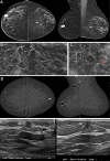
Images in a 77-year-old woman with invasive ductal carcinoma (IDC)
seen only at contrast-enhanced mammography (CEM) and ductal carcinoma in
situ (DCIS) seen only on low-energy (LE) images and digital breast
tomosynthesis images at year 2. (A) Bilateral craniocaudal (CC) (left) and
mediolateral oblique (MLO) (right) LE images show heterogeneously dense
parenchyma and postsurgical changes with dystrophic calcifications in the
upper right breast and clips in the right axilla from breast-conserving
therapy for a grade 1 IDC, estrogen receptor (ER)– and progesterone
receptor (PR)–positive, human epidermal growth factor receptor 2
(HER2) (ERBB2 gene)–negative, 19 years earlier, for which she had
taken Anastrozole for 4 years. Only observer 1 recalled the participant for
linear calcifications in the left breast (arrows), which are better seen on
(B) close-up CC (left) and MLO (right) LE images (arrows) and (C) a spot
magnification CC view of the central left breast. In addition to typically
benign calcifications more laterally and fine linear calcifications (arrow
in C), seen only on C more anteriorly, there is a group of amorphous
calcifications (circle in C). Both areas of calcification were recommended
for biopsy, despite lack of enhancement on (D) recombined CC (left) and MLO
(right) CEM images. A moderately conspicuous enhancing mass in the
retroareolar left breast was newly seen, which was only evident at CC CEM
(arrow in D) and was assessed as Breast Imaging Reporting and Data System
(BI-RADS) 4A, low suspicion, by observer 1 and as BI-RADS 4B, moderate
suspicion, by observer 2. (E) CEM-directed US images (left = transverse
plane, right = longitudinal plane) of the retroareolar left breast show an
irregular, hypoechoic mass (arrows) with an echogenic rim, highly suggestive
of malignancy (BI-RADS 5). US-guided core biopsy and mastectomy revealed a
2.1-cm grade 3 ER and PR-positive, ERBB2-negative IDC. Stereotactic biopsy
of the linear calcifications revealed high-grade ER and PR-positive DCIS,
and biopsy of the amorphous calcifications yielded atypical ductal
hyperplasia. At mastectomy, 5.5 cm of high-nuclear-grade DCIS was found,
discontinuous with the retroareolar IDC. One of two sentinel nodes showed
isolated tumor cells (N0).
Figure 3:
Images in a 77-year-old woman with invasive ductal carcinoma (IDC) seen only at contrast-enhanced mammography (CEM) and ductal carcinoma in situ (DCIS) seen only on low-energy (LE) images and digital breast tomosynthesis images at year 2. (A) Bilateral craniocaudal (CC) (left) and mediolateral oblique (MLO) (right) LE images show heterogeneously dense parenchyma and postsurgical changes with dystrophic calcifications in the upper right breast and clips in the right axilla from breast-conserving therapy for a grade 1 IDC, estrogen receptor (ER)– and progesterone receptor (PR)–positive, human epidermal growth factor receptor 2 (HER2) (ERBB2 gene)–negative, 19 years earlier, for which she had taken Anastrozole for 4 years. Only observer 1 recalled the participant for linear calcifications in the left breast (arrows), which are better seen on (B) close-up CC (left) and MLO (right) LE images (arrows) and (C) a spot magnification CC view of the central left breast. In addition to typically benign calcifications more laterally and fine linear calcifications (arrow in C), seen only on C more anteriorly, there is a group of amorphous calcifications (circle in C). Both areas of calcification were recommended for biopsy, despite lack of enhancement on (D) recombined CC (left) and MLO (right) CEM images. A moderately conspicuous enhancing mass in the retroareolar left breast was newly seen, which was only evident at CC CEM (arrow in D) and was assessed as Breast Imaging Reporting and Data System (BI-RADS) 4A, low suspicion, by observer 1 and as BI-RADS 4B, moderate suspicion, by observer 2. (E) CEM-directed US images (left = transverse plane, right = longitudinal plane) of the retroareolar left breast show an irregular, hypoechoic mass (arrows) with an echogenic rim, highly suggestive of malignancy (BI-RADS 5). US-guided core biopsy and mastectomy revealed a 2.1-cm grade 3 ER and PR-positive, ERBB2-negative IDC. Stereotactic biopsy of the linear calcifications revealed high-grade ER and PR-positive DCIS, and biopsy of the amorphous calcifications yielded atypical ductal hyperplasia. At mastectomy, 5.5 cm of high-nuclear-grade DCIS was found, discontinuous with the retroareolar IDC. One of two sentinel nodes showed isolated tumor cells (N0).
Images in a 47-year-old woman with recurrent invasive lobular
carcinoma (ILC) at the scar detected clinically 6 months after imaging
(interval cancer). (A) Close-up craniocaudal (CC) (left) and mediolateral
oblique (MLO) (right) digital breast tomosynthesis 6-mm slab images of the
right breast show the area of scarring from breast-conserving therapy 2
years earlier for multifocal estrogen receptor (ER)/progesterone receptor
(PR)-positive, human epidermal growth factor receptor 2 (HER2) (ERBB2
gene)–positive ILC, interpreted as benign, Breast Imaging Reporting
and Data System (BI-RADS) 2, by both observers. The participant completed
both neoadjuvant and adjuvant chemotherapy and was taking tamoxifen. (B)
Close-up CC (left) and MLO (right) recombined contrast-enhanced mammography
(CEM) images show nonmass enhancement at the scar (arrows), interpreted as
benign, BI-RADS 2, by both observers. (C) US scan in longitudinal plane
(left) obtained 6 months later, when the participant reported feeling a lump
at the scar, shows a superficial irregular, parallel, hypoechoic mass
(arrow) at the scar; the mass shows internal vascularity on transverse power
Doppler scan (right, arrow). US-guided core biopsy and mastectomy revealed a
2.4-cm grade 3 ILC, ER-positive, PR-negative, HER2-positive lesion (Ki-67
proliferation index of 90%), with one of two sentinel nodes showing isolated
tumor cells (N0).
Figure 4:
Images in a 47-year-old woman with recurrent invasive lobular carcinoma (ILC) at the scar detected clinically 6 months after imaging (interval cancer). (A) Close-up craniocaudal (CC) (left) and mediolateral oblique (MLO) (right) digital breast tomosynthesis 6-mm slab images of the right breast show the area of scarring from breast-conserving therapy 2 years earlier for multifocal estrogen receptor (ER)/progesterone receptor (PR)-positive, human epidermal growth factor receptor 2 (HER2) (ERBB2 gene)–positive ILC, interpreted as benign, Breast Imaging Reporting and Data System (BI-RADS) 2, by both observers. The participant completed both neoadjuvant and adjuvant chemotherapy and was taking tamoxifen. (B) Close-up CC (left) and MLO (right) recombined contrast-enhanced mammography (CEM) images show nonmass enhancement at the scar (arrows), interpreted as benign, BI-RADS 2, by both observers. (C) US scan in longitudinal plane (left) obtained 6 months later, when the participant reported feeling a lump at the scar, shows a superficial irregular, parallel, hypoechoic mass (arrow) at the scar; the mass shows internal vascularity on transverse power Doppler scan (right, arrow). US-guided core biopsy and mastectomy revealed a 2.4-cm grade 3 ILC, ER-positive, PR-negative, HER2-positive lesion (Ki-67 proliferation index of 90%), with one of two sentinel nodes showing isolated tumor cells (N0).

Similar articles

Cited by

References

    1. Lowry KP , Ichikawa L , Hubbard RA , et al. . Variation in second breast cancer risk after primary invasive cancer by time since primary cancer diagnosis and estrogen receptor status . Cancer 2023. ; 129 ( 8 ): 1173 – 1182 . - PMC - PubMed
    1. Houssami N , Ciatto S , Martinelli F , Bonardi R , Duffy SW . Early detection of second breast cancers improves prognosis in breast cancer survivors . Ann Oncol 2009. ; 20 ( 9 ): 1505 – 1510 . - PubMed
    1. Houssami N , Abraham LA , Miglioretti DL , et al. . Accuracy and outcomes of screening mammography in women with a personal history of early-stage breast cancer . JAMA . 2011. ; 305 ( 8 ): 790 – 799 . - PMC - PubMed
    1. Lowry KP , Braunstein LZ , Economopoulos KP , et al. . Predictors of surveillance mammography outcomes in women with a personal history of breast cancer . Breast Cancer Res Treat 2018. ; 171 ( 1 ): 209 – 215 . - PubMed
    1. Yeom YK , Chae EY , Kim HH , Cha JH , Shin HJ , Choi WJ . Screening mammography for second breast cancers in women with history of early-stage breast cancer: factors and causes associated with non-detection . BMC Med Imaging 2019. ; 19 ( 1 ): 2 . - PMC - PubMed

Publication types

LinkOut - more resources