. 2024 Apr 30;147(1):77.
doi: 10.1007/s00401-024-02723-z.

Viral entry and translation in brain endothelia provoke influenza-associated encephalopathy

Affiliations

Viral entry and translation in brain endothelia provoke influenza-associated encephalopathy

Shihoko Kimura-Ohba et al. Acta Neuropathol. .

Abstract

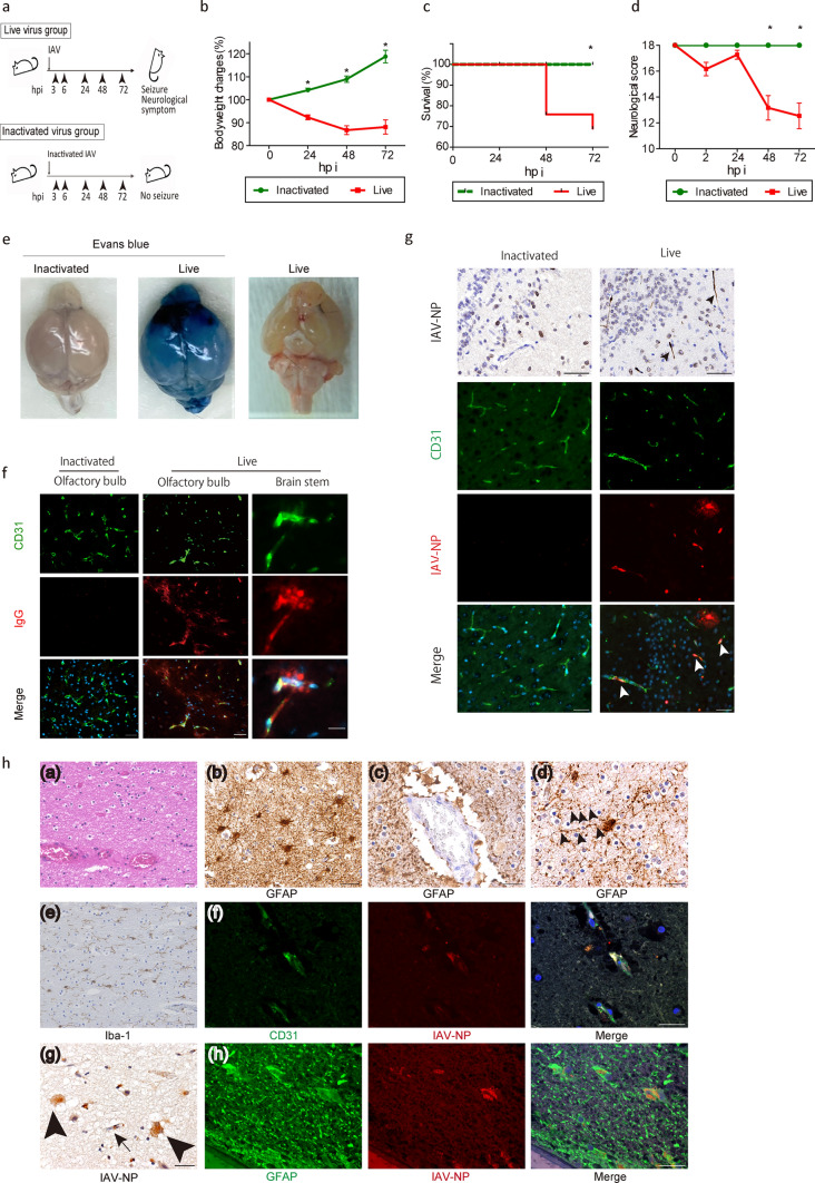
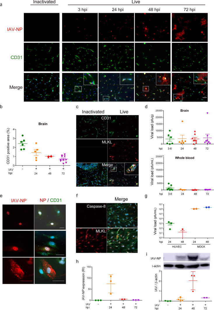
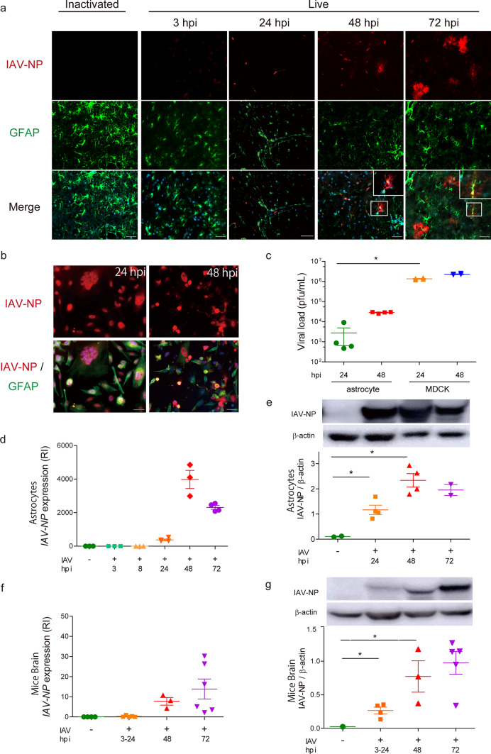
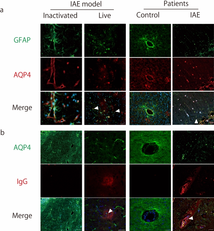
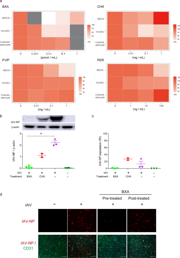
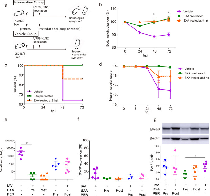
Influenza-associated encephalopathy (IAE) is extremely acute in onset, with high lethality and morbidity within a few days, while the direct pathogenesis by influenza virus in this acute phase in the brain is largely unknown. Here we show that influenza virus enters into the cerebral endothelium and thereby induces IAE. Three-weeks-old young mice were inoculated with influenza A virus (IAV). Physical and neurological scores were recorded and temporal-spatial analyses of histopathology and viral studies were performed up to 72 h post inoculation. Histopathological examinations were also performed using IAE human autopsy brains. Viral infection, proliferation and pathogenesis were analyzed in cell lines of endothelium and astrocyte. The effects of anti-influenza viral drugs were tested in the cell lines and animal models. Upon intravenous inoculation of IAV in mice, the mice developed encephalopathy with brain edema and pathological lesions represented by micro bleeding and injured astrocytic process (clasmatodendrosis) within 72 h. Histologically, massive deposits of viral nucleoprotein were observed as early as 24 h post infection in the brain endothelial cells of mouse models and the IAE patients. IAV inoculated endothelial cell lines showed deposition of viral proteins and provoked cell death, while IAV scarcely amplified. Inhibition of viral transcription and translation suppressed the endothelial cell death and the lethality of mouse models. These data suggest that the onset of encephalopathy should be induced by cerebral endothelial infection with IAV. Thus, IAV entry into the endothelium, and transcription and/or translation of viral RNA, but not viral proliferation, should be the key pathogenesis of IAE.

Keywords: Brain endothelial cells; Clasmatodendrosis; Influenza A virus; Influenza-associated encephalopathy (IAE); Transcription.


Conflict of interest statement

The authors report no competing interests.

Figures

Fig. 1
Fig. 1
Massive deposits of viral protein in the brain of the Influenza virus-induced brain edema (IVE) models and the human IAE patients. a Protocol for inducing IVE. Three weeks-old male C57BL/6 mice were inoculated with Influenza A virus (IAV) A/PR/8/34 at a dose of 1.2 × 103 TCID50/g. Mice were followed up at 3, 8, 24, 48, and 72 h post inoculation (hpi). Control mice were inoculated with inactivated IAV. b Change in body weight, c Kaplan–Meier analysis of survival rate, and d neurological scores of IVE mice, n = 36. e The brains of IVE mice harvested at 72 hpi with (left and middle panels) or (right panel) without injection with Evans blue dye. n = 3. f Representative images of brains from IVE mice harvested at 72 hpi stained with IgG. Red, IgG; green, CD31, a marker of endothelial cells (EC); blue, DAPI for nucleus counterstaining; n = 8. g Representative images of olfactory bulbs from IVE mice harvested at 72 hpi stained with anti-IAV nucleoprotein (NP). Black and white arrowheads indicate IAV-NP positive EC. Red, IAV-NP; green, CD31; blue, DAPI for nucleus counterstaining; n = 5. h Images of the autopsied brain from a patient with IAE. (a) Hematoxylin–Eosin (H&E) staining of the white matter of the frontal cortex, (b, c, d) immunohistochemical staining for GFAP of the cerebral white matter. Arrowheads indicate beaded astrocytic foot processes [(d)]. (e)immunohistochemical staining for Iba-1 of the cerebral white matter, (f) immunohistochemical staining of the cerebral cortex. Red, IAV-NP; green, CD31; blue, DAPI (g) immunohistochemical staining with anti-IAV-NP in the cerebral cortex. An arrow and arrowheads indicate endothelial cell and astrocytes, respectively. (h) immunohistochemistry of the cerebral cortex. Red, IAV-NP; green, a marker of glial fibrillary acidic protein (GFAP); blue, DAPI. Scale bars indicate [f right panels; (f)(h)] 20 μm, (f left and middle panels; g; h [(a), (b), (c), (d), (e), (g)]) 50 μm. Data presented as mean ± SEM, *p < 0.05
Fig. 2
Fig. 2
IAV invades brain endothelial cells and induces necrosis. a Representative images of the thalamus of IVE mice harvested at the indicated hpi stained with anti-IAV-NP antibody. Red, IAV-NP; green, CD31; blue, DAPI, n = 3. b CD31-positive area of brains, n = 4–10 c Images of the brains of IVE mice at 72 hpi stained with an anti-phospho-MLKL antibody. Red, an anti phospho-MLKL (a marker of necroptosis); green, CD31; blue, DAPI, n = 3. d Plaque titration assay of the brain (upper panel) and whole blood (lower panel) harvested from IVE mice at indicated hpi, n = 6–8. e Immunohistochemical analysis of HUVEC at 24 hpi stained with an anti-IAV-NP antibody. Red, an anti-IAV-NP; green, CD31; blue, DAPI, n = 6. f Immunohistochemical analysis of HUVEC at 24 hpi stained with an anti-caspase-8 or an anti-MLKL antibody. Red, caspase-8 (a marker of apoptosis) (upper panel); MIKL (a marker of necroptosis) (lower panel); green, CD31; blue, DAPI, n = 3. g Plaque titration assay of HUVEC or MDCK cells at indicated hpi, n = 3–4. h Expression of the IAV-NP gene and i quantification of IAV-NP by western blotting in HUVEC at the indicated hpi, RI, relative index, n = 3–4. Scale bars indicate (a, c, e, f) 50 μm. Data presented as mean ± SEM, *p < 0.05
Fig. 3
Fig. 3
Infection of IAV in EC precedes viral protein depositions in astrocytes. a Representative images of brains (thalamus, olfactory bulb and cerebellum) from IVE mice harvested at the indicated hpi stained with anti-IAV-NP and anti-GFAP antibody. Green, GFAP (a marker for astrocytes); red; IAV-NP; blue; DAPI, n = 5. b Images of cultured astrocytes in 24 (left panels) and 48 (right panels) hpi stained with an anti-IAV-NP and an anti-GFAP antibodies. Green, GFAP; red, IAV-NP; blue, DAPI, n = 4. c Plaque titration assay of cultured astrocytes and MDCK cells at indicated hpi. n = 24. d Expression of IAV-NP gene and e quantification of IAV-NP in cultured astrocytes at the indicated hpi, RI, relative index, n = 4. f Expression of IAV-NP gene and g quantification of IAV-NP in the brains of IVE mice at the indicated hpi, RI, relative index, n = 3–7. Scale bars indicate a, b 50 μm. Data presented by mean ± SEM, *p < 0.05
Fig. 4
Fig. 4
IAV-NP depositions in the brain EC and astrocytes of the IAE patient. a Upper panels: a lower magnification view (×100) of the cortex from the brain of IAE patients, middle and lower panels: a higher magnification view (×400) of the cortex from the brains of the IAE (middle) and a control (lower) patient. Green, CD31; red, IAV-NP; blue, DAPI. b Staining of the cortex from the brain of the IAE patients. Green, CD31; red, (upper panels) MLKL; or (lower panels) caspase-8; blue, DAPI. Scale bars indicate 50 μm (a middle and lower panels, b) and 400 μm (a upper panels)
Fig. 5
Fig. 5
Endothelial leakage and fragmentation of the astrocytic foot process are observed in the brains of IVE mice and in the human IAE patients. a, b Representative images from the brains of mice inoculated with inactivated IAV (left panels), IVE mice harvested at 72 hpi (live, left middle panels), a human control (right middle panels), and patients with IAE (right panels). a Green, GFAP; red; aquaporin 4 (AQP4); blue, DAPI. Arrowheads indicate fragmented astrocytic processes. b Green, AQP4; red, IgG; blue, DAPI. Arrowheads indicate AQP4-positive fragmented foot process in the IgG leaked area. Scale bars indicate 50 μm
Fig. 6
Fig. 6
Inhibitions of viral transcription and/or translation suppresses endothelial cell death. a The cytotoxicity of IAV-inoculated MDCK, HUVEC and cultured astrocytes treated with Baloxavir acid (BXA), favipiravir (FVP), cycloheximide (CHX), and peramivir trihydrate (PER). Results are shown in heatmap display. Gray box, not tested. n = 3. b Quantification of IAV-NP. c Expression of the IAV-NP gene in IAV-inoculated HUVEC treated with BXA or CHX. RI, relative index; n = 2–3. d Images of IAV-inoculated HUVEC at 48 hpi pre- or post-treated with BXA. Immunocytochemical staining was performed using an anti-IAV-NP and an anti-CD31 antibodies; n = 2–3. Green, CD31; red, IAV-NP; blue, DAPI. Scale bars indicate 50 μm
Fig. 7
Fig. 7
Prominent suppression of lethality in IVE mice by inhibiting viral transcription and translation. a Schematic diagram of the experimental schedule. Male IVE mice were treated with BXA, either before (pre-treatment) or 8 h after inoculation (post-treatment). b Change in body weight, c Kaplan–Meier analysis of the survival rate, and d neurological scores of IVE mice; n = 5–11. e Plaque titration assay of the brains of IVE mice treated with BXA or PER. n = 4–9. f Expression of the IAV-NP gene and g quantification of IAV-NP in brains of IVE mice at 72 hpi treated with BXA or PER. RI, relative index; n = 3–6. Red, IAV-NP; green, CD31; blue, DAPI; n = 3. Scale bars indicate 50 μm. Data presented as means ± SEM, *p < 0.05

References

    1. Amin R, Ford-Jones E, Richardson SE, MacGregor D, Tellier R, Heurter H, et al. Acute childhood encephalitis and encephalopathy associated with influenza: a prospective 11-year review. Pediatr Infect Dis J. 2008;27(5):390–395. doi: 10.1097/INF.0b013e31816507b2. - DOI - PubMed
    1. Armstrong SM, Wang C, Tigdi J, Si X, Dumpit C, Charles S, et al. Influenza infects lung microvascular endothelium leading to microvascular leak: role of apoptosis and claudin-5. PLoS ONE. 2012;7(10):e47323. doi: 10.1371/journal.pone.0047323. - DOI - PMC - PubMed
    1. Azuma J, Nabatame S, Nakano S, Iwatani Y, Kitai Y, Tominaga K, et al. Prognostic factors for acute encephalopathy with bright tree appearance. Brain Dev. 2015;37(2):191–199. doi: 10.1016/j.braindev.2014.04.001. - DOI - PubMed
    1. Balaban D, Miyawaki EK, Bhattacharyya S, Torre M (2021) The phenomenon of clasmatodendrosis. Heliyon 7(7):e07605. 10.1016/j.heliyon.2021.e07605 - PMC - PubMed
    1. Bissel SJ, Giles BM, Wang G, Olevian DC, Ross TM, Wiley CA. Acute murine H5N1 influenza A encephalitis. Brain Pathol. 2012;22(2):150–158. doi: 10.1111/j.1750-3639.2011.00514.x. - DOI - PMC - PubMed

Publication types

MeSH terms

LinkOut - more resources