. 2024 May 24;9(5):2455-2464.
doi: 10.1021/acssensors.4c00153. Epub 2024 Apr 30.

Three-Dimensional Microfluidic Capillary Device for Rapid and Multiplexed Immunoassays in Whole Blood

Affiliations

Three-Dimensional Microfluidic Capillary Device for Rapid and Multiplexed Immunoassays in Whole Blood

Thomas Mortelmans et al. ACS Sens. .

Abstract

In this study, we demonstrate whole blood immunoassays using a microfluidic device optimized for conducting rapid and multiplexed fluorescence-linked immunoassays. The device is capable of handling whole blood samples without any preparatory treatment. The three-dimensional channels in poly(methyl methacrylate) are designed to passively load bodily fluids and, due to their linearly tapered profile, facilitate size-dependent immobilization of biofunctionalized particles. The channel geometry is optimized to allow for the unimpeded flow of cellular constituents such as red blood cells (RBCs). Additionally, to make the device easier to operate, the biofunctionalized particles are pretrapped in a first step, and the channel is dried under vacuum, after which it can be loaded with the biological sample. This novel approach and design eliminated the need for traditionally laborious steps such as filtering, incubation, and washing steps, thereby substantially simplifying the immunoassay procedures. Moreover, by leveraging the shallow device dimensions, we show that sample loading to read-out is possible within 5 min. Our results also show that the presence of RBCs does not compromise the sensitivity of the assays when compared to those performed in a pure buffer solution. This highlights the practical adaptability of the device for simple and rapid whole-blood assays. Lastly, we demonstrate the device's multiplexing capability by pretrapping particles of different sizes, each functionalized with a different antigen, thus enabling the performance of multiplexed on-chip whole-blood immunoassays, showcasing the device's versatility and effectiveness toward low-cost, simple, and multiplexed sensing of biomarkers and pathogens directly in whole blood.

Keywords: blood; diagnostics; immunosensors; microfluidics; multiplexing.


Conflict of interest statement

The authors declare the following competing financial interest(s): The authors declare the following competing interests: EP21171944 and EP2022P04672.

Figures
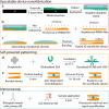
Figure 1
Figure 1
Overview of the 3D nanofluidic immunoassay process. (a) Manufacturing of the master structure through grayscale e-beam lithography in PMMA 950 K, spin-coated on a silicon wafer. The master structure was subject to pattern replication to fabricate a negative daughter stamp for hot embossing. (b) Hot-embossing of a flat PMMA sheet with the negative daughter stamp, followed by UV/ozone-activation at 172 nm. (c) Functionalization of the PMMA surface with PVA through spin-coating and spin-washing. The two PMMA films were aligned and thermally bonded. (d) Functionalization of 2.8 μm particles with BSA via the high-affinity interaction of streptavidin and biotin. Similar functionalization was performed for horseradish peroxidase (HRP) particles. (e) Size-dependent immobilization of biofunctionalized particles and vacuum drying to finalize self-powered patterning of testing lines. (f) One drop immunoassay by mixing the sample under investigation with detection antibodies and loading it into the device through capillary forces.
Figure 2
Figure 2
3D microfluidic device geometry. (a) Schematic showing the different microfluidic components of the capillary device. The top section shows a top view, and the bottom section is a cross-section to highlight the change in channel topography. (b) Bright-field image of a bonded microfluidic device, showing the flow direction as well as the three-dimensional region (3DR). (c) Confocal micrograph of the three-dimensional topography in the 3DR of a device with an outflow height of 800 nm.
Figure 3
Figure 3
On-chip height screening. (a) Profiles of 9 different wedge topographies which were used to find the ideal outflow height. The trapping region indicates where the particle immobilization takes place. The connecting region is representative of the tapering that connects the trapping region with the CP. (b) Photograph showing a PMMA-based chip containing 9 different devices with different channel topographies in the patterned area. Each CP has a width of 2 mm.
Figure 4
Figure 4
Fluorescence images of whole blood flowing through devices with different outflow heights. The channels shown have a width of 2 mm. The investigated dilutions were undiluted, 1:2, and 1:40 in PBS.
Figure 5
Figure 5
Filling velocity of the CP region at different concentrations of whole blood and various outflow heights. The data was fit with a segmental linear regression fit. The two fit functions are given by the solid and dashed black lines.
Figure 6
Figure 6
On-chip fluorescent immunosorbent assays in whole blood. (a) Time lapse showing changes in the fluorescent signal of the trapped particles over time, for both BSA+ and BSA– samples. (b) Fluorescent micrographs of BSA+ and BSA– samples at different times after device loading. The concentration of the secondary C5 donkey antirabbit antibody was 50 nM. The scale bar represents 10 μm.
Figure 7
Figure 7
LOD of immunosorbent assays in whole blood and PBS. (a) Schematic of the immunoassay concept. (b) LOD curve showing the fluorescence intensity for different concentrations of anti-BSA antibody in diluted whole blood (1:40; red dotted line) and PBS (orange dotted line). (c) Bright-field and fluorescent micrographs of biofunctionalized particles in the TR when loaded with different concentrations of anti-BSA antibody in PBS (top) or whole blood (bottom). The RBCs are outlined by a dashed line. The scale bar represents 10 μm.
Figure 8
Figure 8
Multiplexed on-chip immunoassays. (a) Fluorescence images of 2 μm HRP and 2.8 μm biofunctionalized particles in different experimental conditions to show the multiplexing capabilities of the 3D microfluidic device. (b) Quantified fluorescent signal from particles shown in (a). The error bars represent the standard error of the mean (N = 3). All the scale bars represent 10 μm.

Similar articles

References

    1. Hage D. S. Immunoassays. Anal. Chem. 1999, 71, 294R–304R. 10.1021/a1999901+. - DOI - PubMed
    1. Pinho A. R.; Fortuna A.; Falcão A.; Santos A. C.; Seiça R.; Estevens C.; Veiga F.; Ribeiro A. J. Comparison of ELISA and HPLC-MS Methods for the Determination of Exenatide in Biological and Biotechnology-Based Formulation Matrices. J. Pharm. Anal. 2019, 9 (3), 143–155. 10.1016/j.jpha.2019.02.001. - DOI - PMC - PubMed
    1. de Bruin E.; Duizer E.; Vennema H.; Koopmans M. P. G. Diagnosis of Norovirus Outbreaks by Commercial ELISA or RT-PCR. J. Virol. Methods 2006, 137 (2), 259–264. 10.1016/j.jviromet.2006.06.024. - DOI - PubMed
    1. Ng A. H. C.; Uddayasankar U.; Wheeler A. R. Immunoassays in Microfluidic Systems. Anal. Bioanal. Chem. 2010, 397 (3), 991–1007. 10.1007/s00216-010-3678-8. - DOI - PubMed
    1. Kohl T. O.; Ascoli C. A. Immunometric Double-Antibody Sandwich Enzyme-Linked Immunosorbent Assay. Cold Spring Harb. Protoc. 2017, 2017 (6), pdb.prot093724.10.1101/pdb.prot093724. - DOI - PubMed

Publication types

MeSH terms

Substances