Molecular and clinical presentation of UBA1-mutated myelodysplastic syndromes
- PMID: 38687605
- PMCID: PMC12060156
- DOI: 10.1182/blood.2023023723
Molecular and clinical presentation of UBA1-mutated myelodysplastic syndromes
Abstract
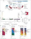
Mutations in UBA1, which are disease-defining for VEXAS (vacuoles, E1 enzyme, X-linked, autoinflammatory, somatic) syndrome, have been reported in patients diagnosed with myelodysplastic syndromes (MDS). Here, we define the prevalence and clinical associations of UBA1 mutations in a representative cohort of patients with MDS. Digital droplet polymerase chain reaction profiling of a selected cohort of 375 male patients lacking MDS disease-defining mutations or established World Health Organization (WHO) disease classification identified 28 patients (7%) with UBA1 p.M41T/V/L mutations. Using targeted sequencing of UBA1 in a representative MDS cohort (n = 2027), we identified an additional 27 variants in 26 patients (1%), which we classified as likely/pathogenic (n = 12) and of unknown significance (n = 15). Among the total 40 patients with likely/pathogenic variants (2%), all were male and 63% were classified by WHO 2016 criteria as MDS with multilineage dysplasia or MDS with single-lineage dysplasia. Patients had a median of 1 additional myeloid gene mutation, often in TET2 (n = 12), DNMT3A (n = 10), ASXL1 (n = 3), or SF3B1 (n = 3). Retrospective clinical review, where possible, showed that 82% (28/34) UBA1-mutant cases had VEXAS syndrome-associated diagnoses or inflammatory clinical presentation. The prevalence of UBA1 mutations in patients with MDS argues for systematic screening for UBA1 in the management of MDS.
© 2024 American Society of Hematology. Published by Elsevier Inc. All rights are reserved, including those for text and data mining, AI training, and similar technologies.
Conflict of interest statement
Conflict-of-interest disclosure: U.G. has received honoraria from Celgene, Novartis, Amgen, Janssen, Roche, and Jazz; and has received research funding from Celgene and Novartis. A.A.v.d.L. serves on advisory boards of Celgene, Amgen, Roche, Novartis, and Alexion; and has received research funding from Celgene. F.T. serves on the advisory boards of Jazz, Pfizer, and AbbVie; and has received research funding from Celgene. I.K. serves on the advisory board of Genesis Pharma; and has received research funding from Celgene and Janssen Hellas. F.P.S.S. has received honoraria from Janssen-Cilag, Bristol Myers Squibb, Novartis, Amgen, AbbVie, and Pfizer; serves on the advisory boards of Novartis, Amgen, and AbbVie; and has received research funding from Novartis. M.R.S. serves on the advisory boards of AbbVie, Astex, Celgene, Karyopharm, Selvita, and TG Therapeutic; has equity in Karyopharm; and has received research funding from Astex, Incyte, Sunesis, Takeda, and TG Therapeutics. G.S. serves on the advisory boards of AbbVie, Amgen, Astellas, Böehringer-Ingelheim, Celgene, Helsinn Healthcare, Hoffmann-La Roche, Janssen-Cilag, Novartis, and Onconova; and has received research funding from Celgene, Hoffmann-La Roche, Janssen-Cilag, and Novartis. L.A. serves on the advisory boards of AbbVie, Astex, Celgene, and Novartis and has received research funding from Celgene. M.H. has received honoraria from Novartis, Pfizer, and PriME Oncology; serves on the advisory boards of AbbVie, Bayer Pharma, Daiichi Sankyo, Novartis, and Pfizer and has received institutional research funding from Astellas, Bayer Pharma, BergenBio, Daiichi Sankyo, Karyopharm, Novartis, Pfizer, and Roche. P.V. has received honoraria and research funding from Celgene. C.F. serves on the advisory boards of, and has received honoraria from, Celgene, Novartis, and Janssen; and has received research funding from Celgene. M.T.V. serves on the advisory board of Celgene; has received honoraria from Celgene and Novartis; and has received research funding from Celgene. N.G. serves on the advisory board of, and has received honoraria from, Novartis; and has received research funding from Alexion. B.L.E. has received research funding from Celgene and Deerfield. R.B. serves on the advisory boards of Celgene, AbbVie, Astex, NeoGenomics, and Daiichi Sankyo; and has received research funding from Celgene and Takeda. D.B.B. receives consulting fees from Alexion Pharmaceuticals and is on the advisory boards for Novartis and Sobi. E.H.-L. has received research funding from Celgene. E.P. is a founder, equity holder, and holds fiduciary roles in Isabl Inc, a company offering analytics for cancer whole-genome sequencing data; and holds stock options in TenSixteen Bio. The remaining authors declare no competing financial interests.
Figures


Comment in
-
Bone marrow vexations.Blood. 2024 Sep 12;144(11):1140-1141. doi: 10.1182/blood.2024024971. Blood. 2024. PMID: 39264609 No abstract available.
References
-
- Bernard E, Tuechler H, Greenberg PL, et al. Molecular international prognostic scoring system for myelodysplastic syndromes. NEJM Evid. 2022;1(7) - PubMed
-
- Braun T, Fenaux P. Myelodysplastic syndromes (MDS) and autoimmune disorders (AD): cause or consequence? Best Pract Res Clin Haematol. 2013;26(4):327–336. - PubMed
MeSH terms
Substances
Grants and funding
LinkOut - more resources
Full Text Sources
Medical
Molecular Biology Databases
Research Materials
Miscellaneous

