. 2024 Apr 30;134(11):e168649.
doi: 10.1172/JCI168649.

Androgen receptor splice variants drive castration-resistant prostate cancer metastasis by activating distinct transcriptional programs

Affiliations

Androgen receptor splice variants drive castration-resistant prostate cancer metastasis by activating distinct transcriptional programs

Dong Han et al. J Clin Invest. .

Abstract

One critical mechanism through which prostate cancer (PCa) adapts to treatments targeting androgen receptor (AR) signaling is the emergence of ligand-binding domain-truncated and constitutively active AR splice variants, particularly AR-V7. While AR-V7 has been intensively studied, its ability to activate distinct biological functions compared with the full-length AR (AR-FL), and its role in regulating the metastatic progression of castration-resistant PCa (CRPC), remain unclear. Our study found that, under castrated conditions, AR-V7 strongly induced osteoblastic bone lesions, a response not observed with AR-FL overexpression. Through combined ChIP-seq, ATAC-seq, and RNA-seq analyses, we demonstrated that AR-V7 uniquely accesses the androgen-responsive elements in compact chromatin regions, activating a distinct transcription program. This program was highly enriched for genes involved in epithelial-mesenchymal transition and metastasis. Notably, we discovered that SOX9, a critical metastasis driver gene, was a direct target and downstream effector of AR-V7. Its protein expression was dramatically upregulated in AR-V7-induced bone lesions. Moreover, we found that Ser81 phosphorylation enhanced AR-V7's pro-metastasis function by selectively altering its specific transcription program. Blocking this phosphorylation with CDK9 inhibitors impaired the AR-V7-mediated metastasis program. Overall, our study has provided molecular insights into the role of AR splice variants in driving the metastatic progression of CRPC.

Keywords: Molecular genetics; Oncology; Prostate cancer; Transcription.


Figures
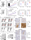
Figure 1
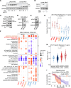
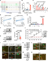
Figure 1. Overexpression of AR-V7, but not AR-FL, induces osteoblastic bone metastasis in PCa.
(A and B) Immunoblotting for AR-V7 or AR-FL in C4-2–derived lentiviral stable lines overexpressing doxycycline-regulated, V5-tagged AR-V7 (C4-2-tet-ARV7) (A) or AR-FL (C4-2-tet-ARFL) (B). Cells were pretreated with or without 0.25 μg/mL doxycycline for 48 hours. (C and D) C4-2-tet-ARV7 (C) or C4-2-tet-ARFL (D) cells were injected into the tibias of castrated NSG mice, which were then fed with or without a doxycycline-supplemented diet. The bone lesion area was monitored and quantified. (E and F) Normalized bone volume (BV) and trabecular bone number (Tb.N) in C4-2-tet-ARV7 tumors (E) or C4-2-tet-ARFL tumors (F) were compared. TV, total volume. (G) Structural views of bones scanned by μCT and 3D reconstructed for the C4-2-tet-ARV7 model. (H and I) Immunohistochemistry (IHC) staining for SOX9 in tumor samples from the C4-2-tet-ARV7 model (H) and the C4-2-tet-ARFL model (I). Scale bars (H and I): 500 μm (left) and 100 μm (right). (J and K) Immunoblotting for AR (antibody against N-terminus) and SOX9 in C4-2-tet-ARV7 (J) and C4-2-tet-ARFL cells (K), which were treated with 0, 0.1, 0.5, or 1 μg/mL doxycycline for 48 hours. All the cell lines were hormone depleted prior to the experiments. Unpaired, 2-sided Student’s t test was used to determine statistical significance. *P < 0.05, **P < 0.01, ***P < 0.001.
Figure 2
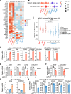
Figure 2. AR-V7 activates a unique transcription program in CRPC.
(A) Immunoblotting for AR (N-terminus) in LNCaP cells stably expressing doxycycline-regulated V5-tagged AR-FL (LN-tet-ARFL) or AR-V7 (LN-tet-ARV7) treated with 0–1 μg/mL doxycycline for 48 hours. (B) Immunoblotting for AR in LN-tet-ARV7 cells treated with or without low-dose doxycycline (0.1 μg/mL for 48 hours) or DHT (10 nM for 24 hours). (C) Immunoblotting for indicated proteins in LNCaP cells stably expressing cumate-regulated FLAG-tagged ARv567es (LN-cu-ARv567es) with the treatment of 0, 30, or 60 μg/mL cumate for 48 hours. (D and E) Immunoblotting for AR-V7 and N-terminal AR in CWR-22Rv1 (D) and LuCaP 35CR cells (E) transfected with siRNAs against nontarget control (NTC) or AR-V7 for 3 days. (F) RNA-seq analyses were conducted to compare the AR-V7 transcriptome (22Rv1 and 35CR transfected with siNTC or siARV7 for 3 days, LN-tet-ARV7 treated with or without 0.25 μg/mL doxycycline for 48 hours) with the ARv567es transcriptome (LN-cu-ARv567es treated with or without 30 μg/mL cumate for 48 hours) and DHT-stimulated AR-FL transcriptome (LNCaP/C4-2/VCaP stimulated with or without 10 nM DHT for 24 hours, LN-tet-ARFL treated with 0.25 μg/mL doxycycline and stimulated with or without 0.1 nM DHT for 24 hours). GSEA normalized enrichment scores (NES) of MSigDB Hallmark gene sets in each model were plotted (red, AR-V7– or AR-FL–activated pathways; blue, AR-V7– or AR-FL–repressed pathways). All the cell lines were hormone depleted prior to the experiments. (G and H) The expression of AR-V7–specific targets (17-gene signature) in these cell lines (G) and in human PCa cohorts: Normal (n = 52) and androgen-dependent primary PCa samples (n = 498) from TCGA data set versus metastatic CRPC samples from SU2C (n = 266) and UW data sets (n = 138) (H). P values are shown above the horizontal bars. (I) Kaplan-Meier survival analysis for the overall survival from the initiation of the first-line ARSi in mCRPC patients (SU2C cohort, n = 99) was conducted, comparing top 25th percentile of median score expression (red, n = 25) versus lower 75th percentile (blue, n = 74). P value was calculated using the log-rank test from the score test. Statistical analyses were conducted using unpaired, nonparametric 2-sample Wilcoxon’s test for box-and-whisker plots, with Bonferroni’s correction for multiple comparisons.
Figure 3
Figure 3. AR-V7–activated transcription program is enriched for EMT/metastasis functions.
(A) Heatmap view of identified AR-V7–activated EMT genes (37-gene signature). (B) GSEA NES of 2 public bone metastasis signatures (VICENT_BONE_MET, 20-gene; CAI_BONE_MET, 44-gene) in each model were plotted (red, activated pathways; blue, repressed pathways). Statistical analyses were conducted using unpaired, nonparametric 2-sample Wilcoxon’s test for box-and-whisker plots, with Bonferroni’s correction for multiple comparisons. (C) Fold-change of AR-V7–activated EMT genes (37-gene signature) in 22RV1, 35CR, LN-tet-ARV7, and LN-tet-ARFL cells. P values by Wilcoxon’s test are shown above the horizontal bars. (D) qRT-PCR for a panel of AR-V7–regulated genes in LN-tet-ARV7 cells treated with or without 0.25 μg/mL doxycycline. (E and F) qRT-PCR for a panel of AR-V7–regulated genes in 22Rv1 (E) or 35CR (F) cells transfected with siNTC or siARV7. (G) qRT-PCR for SOX9 and COL25A1 in LN-cu-ARv567es cells treated with 10 nM DHT for 24 hours or 30 μg/mL cumate for 48 hours. (H) qRT-PCR for a panel of AR-V7–regulated genes in parental LNCaP cells treated with or without 10 nM DHT for 24 hours. All the cell lines were hormone depleted prior to the experiments. For the bar graphs, an unpaired, 2-sided Student’s t test was used to determine statistical significance. *P < 0.05, **P < 0.01, ***P < 0.001, ****P < 0.0001. Data are represented as mean ± SEM.
Figure 4
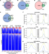
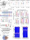
Figure 4. AR-V7 can bind to a subset of chromatin sites distinct from AR-FL binding.
(A and B) ChIP-seq analysis of V5 was conducted in LN-tet-ARV7 cells (hormone depleted) stimulated with or without 0.25 μg/mL doxycycline for 48 hours. Similarly, ChIP-seq analysis of AR (antibody against N-terminus) was performed in LN-tet-ARFL cells stimulated with 0.25 μg/mL doxycycline and then treated with 0.1 nM DHT for 4 hours. The Venn diagram (A) and heatmap view (B) demonstrate the unique or overlapping sites of AR-V7 versus AR-FL. (CE) Binding and expression target analysis (BETA) was used to assess the association of total AR-V7 sites with AR-V7–regulated genes (C), total AR-FL sites with androgen-upregulated genes (D), and the unique or common sites of AR-FL/AR-V7 with androgen-upregulated genes or AR-V7–regulated genes (E). P values were calculated by BETA as a measure of the significance of the association between transcription factor binding and gene expression changes. (F) Motif enrichment analyses were conducted for the AR-FL/AR-V7 unique or common sites and ranked by z score (black, common enriched motifs; purple, uniquely enriched motifs).
Figure 5
Figure 5. AR-V7 can bind to more compact chromatin regions.
(AD) ChIP-seq analyses of FOXA1 were conducted in LN-tet-AR-V7 cells treated with or without 0.25 μg/mL doxycycline for 48 hours. Venn diagrams demonstrate FOXA1 binding sites in treated versus untreated cells (A), FOXA1 binding sites in untreated cells versus AR-V7 sites (B), FOXA1 binding sites in untreated parental LNCaP cells versus AR-FL binding sites (C), and FOXA1 binding sites in doxycycline-treated cells versus AR-V7 sites (D). (E) ATAC-seq and ChIP-seq analyses of H3K27ac were performed in LN-tet-AR-V7 cells treated with or without 0.25 μg/mL doxycycline for 48 hours. The heatmap shows peak intensity of FOXA1, ATAC, and H3K27ac at AR-FL/AR-V7 common sites or AR-V7–specific sites. (FH) The average intensity curves of FOXA1 (F), ATAC (G), and H3K27ac (H) at AR-FL/AR-V7 common sites versus AR-V7–specific sites. All the cell lines were hormone depleted prior to the experiments.
Figure 6
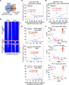
Figure 6. AR-V7 transcriptionally activates SOX9.
(A) Genome browser view for indicated protein binding at the S2 site of the SOX9 gene (Note: AR-FL binding indicates AR ChIP-seq peaks in LN-tet-ARFL cells treated with doxycycline; other tracks are for LN-tet-ARV7 cells). (B) qRT-PCR for SOX9 mRNA in LN-tet-ARFL cells (0.1 nM DHT for 24 hours, 0.25 μg/mL doxycycline for 48 hours) and in LN-tet-ARV7 (0.25 μg/mL doxycycline for 48 hours). (C) ChIP-qPCR for V5 (AR-V7), FOXA1, H3K4me2, H3K27ac, and C-terminal AR (AR-FL) at the S2 site in LN-tet-ARV7 cells treated with/out 0.25 μg/mL doxycycline for 48 hours or 0.1 nM DHT for 4 hours. (D) Spearman’s correlation of SOX9 expression with AR-V7 or AR-FL expression in the SU2C mCRPC data set. (E) Matrigel invasion assay in LN-tet-ARV7 cells (doxycycline) compared with LN-tet-ARFL cells (0–10 nM DHT, 0.1 μg/mL doxycycline for 3 days). (F and G) Immunoblotting for SOX9 (F) and Matrigel invasion assay (G) in LN-tet-ARV7 cells transfected with siNTC or siSOX9 for 3 days. (H) Immunoblotting for SOX9 in GFP-labeled C4-2-tet-ARV7 cells transfected with siNTC or siSOX9 for 3 days. (I) GFP-labeled C4-2-tet-ARFL (grown under 0.1 nM DHT) or C4-2-tet-ARV7 stable cells, pretreated with or with out 0.25 μg/mL doxycycline and transfected with siNTC or siSOX9 for 3 days, were injected into the zebrafish embryos. AR-V7–mediated tumor cell intravasation process was observed within 1 hour (indicated by red arrow). The proportion of invaded embryos relative to the total number of embryos injected is displayed. (J) C4-2 cells stably expressing doxycycline-regulated AR-V7 together with doxycycline-regulated shRNA against SOX9 (LN-tet-ARV7/shSOX9) were established. Immunoblotting for AR-V7 and SOX9 was performed (right panel). LN-tet-ARV7/shSOX9 or control LN-tet-ARV7 cells were then injected into the tibias of castrated male mice, which were then fed with a doxycycline-supplemented diet. The bone lesion area was monitored and quantified (left panel). (K and L) Immunoblotting for AR-V7 and SOX9 (K) and zebrafish embryo metastasis assay (L) in GFP-labeled LNCaP-95 cells transfected with siNTC or siARV7 for 3 days. (M and N) Immunoblotting for AR-V7 and SOX9 (M) and zebrafish embryo metastasis assay (N) in GFP-labeled 35CR cells transfected with siNTC or siARV7 for 3 days. All the cell lines were hormone depleted prior to the experiments. ***P < 0.001; ****P < 0.0001 by unpaired, 2-sided Student’s t test (B, C, E, G, and J), Fisher’s exact test (K and L), or χ2 test (N). Data are represented as mean ± SD.
Figure 7
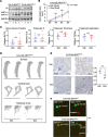
Figure 7. Ser81 phosphorylation is required for AR-V7–induced metastasis.
(A) Immunoblotting for indicated proteins in C4-2-tet-ARV7 and C4-2-ARV7S81A cells (C4-2 cells expressing doxycycline-regulated V5-tagged AR-V7-S81A mutant) treated with 0–1 μg/mL doxycycline for 48 hours. (B) C4-2-ARV7S81A cells were injected into the tibias of castrated male mice, which were then fed with or without a doxycycline-supplemented diet. The bone lesion area was monitored and quantified. Note: this experiment was conducted simultaneously with the C4-2-tet-ARV7 and C4-2-tet-ARFL experiments shown in Figure 1. (C) Normalized bone volume and trabecular bone number were compared. (D) Structure views of bones scanned by μCT and 3D reconstructed. (E) IHC staining for SOX9 in tumor samples. Scale bars: 500 μm (left) and 100 μm (right). (F) Zebrafish embryo metastasis assay in GFP-labeled C4-2-tet-ARV7WT and C4-2-tet-ARV7S81A cells pretreated with or without 0.25 μg/mL doxycycline for 48 hours. All the cell lines were hormone depleted before experiments. **P < 0.01; ***P < 0.001 by unpaired, 2-sided Student’s t test (B, C, and E) or Fisher’s exact test (F).
Figure 8
Figure 8. Ser81 phosphorylation selectively enhances the AR-V7–regulated metastasis program.
(A) Immunoblotting for S81-phosphorylated (p-S81) AR-FL and AR-V7 in LN-tet-ARV7 and LN-tet-ARV7S81A cells (LNCaP cells expressing doxycycline-regulated V5-tagged AR-V7-S81A mutant). (B and C) RNA-seq analyses were conducted in these stable lines treated with or without 0.25 μg/mL doxycycline. GSEA using Hallmark gene sets (B) or predefined bone metastasis gene sets (C) was performed. (D) Relative fold change for AR-V7 regulation of indicated gene sets. (E) qRT-PCR for AR-V7–activated EMT/metastasis genes and lipid synthesis genes. (F) Immunoblotting for SOX9 and AR in LN-tet-ARFL, LN-tet-ARFLS81A, LN-tet-ARV7, and LN-tet-ARV7S81A cells, treated with 10 nM DHT or 0.25 μg/mL doxycycline. (GI) ChIP-seq analysis of V5 was performed in LN-tet-ARV7S81A cells stimulated with or without 0.25 μg/mL doxycycline. The Venn diagram for AR-V7-WT bindings sites versus AR-V7-S81A binding sites (G), heatmap view for peak intensity at AR-V7-WT and AR-V7-S81A unique or common sites (H), and heatmap view for peak intensity at AR-V7 and AR-FL unique or common sites (I) are shown. All the cell lines were hormone depleted before experiments. *P < 0.05; **P < 0.01; ***P < 0.001; ****P < 0.0001 by Wilcoxon’s test (D) or unpaired, 2-sided Student’s t test (E). Data are represented as mean ± SD.
Figure 9
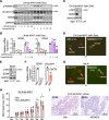
Figure 9. CDK9 inhibition prevents Ser81 phosphorylation of AR-V7 and impairs AR-V7–mediated metastasis.
(A) Immunoblotting for p-S81 of AR-V7, V5, and SOX9 in LN-tet-ARV7 cells treated with CDK9 inhibitors for 24 hours. (B) qRT-PCR for AR-V7 target genes in LN-tet-ARV7 cells treated with 2 CDK9 inhibitors for 24 hours. (C and E) Immunoblotting for p-S81 of AR-V7 and SOX9 in C4-2-tet-ARV7 (C) and 35CR (E) cells treated with or without 0.1 μM AZD4573 for 24 hours. (D and G) Zebrafish embryo metastasis assays in GFP-labeled C4-2-tet-ARV7 cells cultured under doxycycline (D) and 35CR cells (G) pretreated with or without 0.1 μM AZD4573 for 24 hours. (F) qRT-PCR for AR-V7 target genes in 35CR cells treated with 0.1 μM AZD4573 for 24 hours. (H) C4-2-tet-ARV7 cells were injected into the tibias of castrated male mice, which were then fed with a doxycycline-supplemented diet and treated with (n = 12) or without AZD4573 (n = 8, 15 mg/kg) via i.p. injection every other day. The bone lesion area was monitored and quantified. (I) IHC staining for SOX9 in tumor samples. Scale bars: 100 μm. All the cell lines were hormone depleted before experiments. *P < 0.05; **P < 0.01; ***P < 0.001; ****P < 0.0001 by unpaired, 2-sided Student’s t test (bar graphs) or χ2 test (D and G). Data are represented as mean ± SD.

References

    1. Green SM, et al. Androgen action and metabolism in prostate cancer. Mol Cell Endocrinol. 2012;360(1-2):3–13. doi: 10.1016/j.mce.2011.09.046. - DOI - PMC - PubMed
    1. Yuan X, et al. Androgen receptor functions in castration-resistant prostate cancer and mechanisms of resistance to new agents targeting the androgen axis. Oncogene. 2014;33(22):2815–2825. doi: 10.1038/onc.2013.235. - DOI - PMC - PubMed
    1. de Bono JS, et al. Abiraterone and increased survival in metastatic prostate cancer. N Engl J Med. 2011;364(21):1995–2005. doi: 10.1056/NEJMoa1014618. - DOI - PMC - PubMed
    1. Scher HI, et al. Increased survival with enzalutamide in prostate cancer after chemotherapy. N Engl J Med. 2012;367(13):1187–1197. doi: 10.1056/NEJMoa1207506. - DOI - PubMed
    1. Hoang DT, et al. Androgen receptor-dependent and -independent mechanisms driving prostate cancer progression: Opportunities for therapeutic targeting from multiple angles. Oncotarget. 2017;8(2):3724–3745. doi: 10.18632/oncotarget.12554. - DOI - PMC - PubMed

MeSH terms