. 2024 Apr 30;19(4):e0302851.
doi: 10.1371/journal.pone.0302851. eCollection 2024.

The barrier-protective effect of β-eudesmol against type 2-inflammatory cytokine-induced tight junction disassembly in airway epithelial cells

Affiliations

The barrier-protective effect of β-eudesmol against type 2-inflammatory cytokine-induced tight junction disassembly in airway epithelial cells

Phuntila Tharabenjasin et al. PLoS One. .

Abstract

Allergic inflammation, which is the pathogenesis of allergic rhinitis and asthma, is associated with disruption of the airway epithelial barrier due to the effects of type 2 inflammatory cytokines, i.e. interleukin-4 and interleukin-13 (IL-4/13). The anti-allergic inflammatory effect of β-eudesmol (BE) on the tight junction (TJ) of the airway epithelium has not previously been reported. Herein, the barrier protective effect of BE was determined by measurement of transepithelial electrical resistance and by paracellular permeability assay in an IL-4/13-treated 16HBE14o- monolayer. Pre-treatment of BE concentration- and time- dependently inhibited IL-4/13-induced TJ barrier disruption, with the most significant effect observed at 20 μM. Cytotoxicity analyses showed that BE, either alone or in combination with IL-4/13, had no effect on cell viability. Western blot and immunofluorescence analyses showed that BE inhibited IL-4/13-induced mislocalization of TJ components, including occludin and zonula occludens-1 (ZO-1), without affecting the expression of these two proteins. In addition, the mechanism of the TJ-protective effect of BE was mediated by inhibition of IL-4/13-induced STAT6 phosphorylation, in which BE might serve as an antagonist of cytokine receptors. In silico molecular docking analysis demonstrated that BE potentially interacted with the site I pocket of the type 2 IL-4 receptor, likely at Asn-126 and Tyr-127 amino acid residues. It can therefore be concluded that BE is able to prevent IL-4/13-induced TJ disassembly by interfering with cytokine-receptor interaction, leading to suppression of STAT6-induced mislocalization of occludin and ZO-1. BE is a promising candidate for a therapeutic intervention for inflammatory airway epithelial disorders driven by IL-4/13.


Conflict of interest statement

The authors have declared that no competing interests exist.

Figures
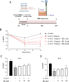
Fig 1
Fig 1. The preventive effect of BE on IL-4/13-induced epithelial barrier disruption in 16HBE14o- cells.
(A) Schematic diagram of barrier integrity measurement. The TEER of the indicated treatment was obtained from pretreatment of cells with vehicle (0.25% v/v DMSO) or BE at dose of 5, 10, and 20 μM prior to exposure to IL-4/13 (10 ng/ml each). (B) Sequential change of TEER after each treatment. (C–D) Observations for the TEER after 24 h and 48 h, respectively. The data represents the mean percentage of changed TEER to baseline ± S.E.M (n = 5–8). Differences in mean values over time were analyzed using two-way ANOVA and Tukey’s post-hoc analysis in Fig 1B, and one-way ANOVA with Dunnett’s post-hoc analysis in Fig 1C and 1D. * P < 0.05,** P < 0.01, *** P < 0.001 compared with control group (black bar); P < 0.05 compared with vehicle-pretreated group (gray bar).
Fig 2
Fig 2. The effect of BE on barrier integrity and tight junction assembly determined by permeability assay.
(A) Schematic diagram of the permeability assay. After 48 h exposure to the indicated treatment, 4-kDa-FITC-dextran (B) or 10-kDa-Texas-red-dextran (C) was added to the apical chamber. The fluorescence intensity of FITC- or Texas-red conjugated dextran leaking from the upper to the lower chambers of the Transwell membranes was measured by sampling media from the basolateral chamber at 2 h. Data are expressed as means of relative fluorescent unit (RFU) of control ± S.E.M (n = 5–6). *** P < 0.001 compared with control group (black bar); P < 0.05 and ††† P < 0.001 compared with the vehicle-pretreated group (gray bar) (one-way ANOVA with Bonferroni multiple comparison test).
Fig 3
Fig 3. Combining effects of BE and IL-4/13 at different concentrations on 16HBE14o- cell survival.
Cell viability was determined by MTT assay after treatment with BE of various concentrations (5–20 μM) alone (A) or in combination with IL-4/13 (each at 10 ng/mL) (B) for 48 h. (C) TEER was measured over time after a prolonged 72 h exposure to BE. The results are expressed as mean ± SEM (n = 4). *** P < 0.001 compared with control group (black bar) (one-way ANOVA); ns: non-significant P-value.
Fig 4
Fig 4. Barrier protective effects of BE might not be via alteration of TJ protein expression in 16HBE14o- cells.
(A) Representative electrophoretic bands of the TJ proteins, ZO-1, and occludin (B) The densitometry analyses of TJ proteins in cells treated for 48 h with IL-4/13 (10 ng/mL each) alone or pre-exposure to 20 μM BE was analyzed by western blot analysis. The histogram bar represents the mean of the ratio of the control ± SEM to the three independent experiments. The values were normalized to beta-actin. * P < 0.05 compared with the control group (black bar) (one-way ANOVA); ns: non-significant P-value.
Fig 5
Fig 5. Immunofluorescence staining of TJ proteins.
(A) Localization of occludin and ZO-1 labeled by immunofluorescence in monolayers treated with IL-4/13 (10 ng/mL each) (Middle panel) or the cytokine plus 20 μM BE (Right panel). Scale bars represent 25 μm. The regions of occludin and ZO-1 were marked and segmented, and intensities of ZO-1 that localized in the occludin region, as well as the occludin in the ZO-1 region, were calculated. The violin plot shows the mean percentage of change in the aforementioned intensities (Right panel). (B) The high magnification of tight junction images in X-Y and X-Z projection (scale bars represent 10 μm). The yellow arrowhead represents internalization of ZO-1 into cytoplasmic sites at 48 h after IL-4/13 treatment. ** P < 0.01 compared with control group; P < 0.05, †† P < 0.01 compared with vehicle-pretreated group (one-way ANOVA).
Fig 6
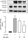
Fig 6. BE suppresses TJ disruption induced by cytokines through the inhibition of STAT6 phosphorylation.
The western blot analyses of STAT6 and the phosphorylated form of STAT6 (pSTAT6) protein were derived from cell extracts after 48 h of the indicated treatment. Data is represented as the mean of the ratio of the control ± SEM of the three independent experiments. Values were normalized to STAT6. *** P < 0.001 compared with control group (black bar); †† P < 0.01 compared with vehicle-pretreated group (gray bar) (one-way ANOVA).
Fig 7
Fig 7. Representation of molecular docking between BE and site I binding pocket of type 2 IL-4R.
(Left) 3D visualization of the four active pockets of the IL-4R; (Right) 3D visualization of BE (yellow structure) in site I pocket of IL-4R. BE was docked to the Asn-126 and Tyr-127 in > 70% of 200 runs and the orientation of BE at the site I pocket, near the Glu-12 of IL-13 (some residues were omitted for clarity).
Fig 8
Fig 8. Proposed underpinning mechanism of BE counteracts IL-4/13 signaling to maintain TJ integrity.
BE selectively blocks type 2 IL-4R at the site I binding pocket, which can interrupt cytokine actions. This inhibition leads to reduction of STAT6, which induce TJ internalization. Of note, STAT6 possibly regulates TJ proteins through alteration of the cytoskeletal architecture, filamentous actin (F-actin) which contributes to internalization of occludin, and ZO-1. The inhibitory effects of BE (cross sign symbol) on IL-4/13 signaling would therefore suppress TJ disruption.

Similar articles

References

    1. Akdis CA. Therapies for allergic inflammation: refining strategies to induce tolerance. Nat Med. 2012; 18(5):736–49. doi: 10.1038/nm.2754 - DOI - PubMed
    1. Gauvreau GM, El-Gammal AI, O’Byrne PM. Allergen-induced airway responses. Eur Respir J. 2015; 46(3):819–31. doi: 10.1183/13993003.00536-2015 - DOI - PubMed
    1. Georas SN, Rezaee F Epithelial barrier function: at the front line of asthma immunology and allergic airway inflammation J Allergy Clin Immunol. 2014; 134(3):509–20. doi: 10.1016/j.jaci.2014.05.049 - DOI - PMC - PubMed
    1. Vareille M, Kieninger E, Edwards MR, Regamey N. The airway epithelium: soldier in the fight against respiratory viruses. Clin Microbiol Rev 2011; 24(1):210–29. doi: 10.1128/CMR.00014-10 - DOI - PMC - PubMed
    1. Noureddine N, Chalubinski M, Wawrzyniak P. The role of defective epithelial barriers in allergic lung disease and asthma development. J Asthma Allergy.2022; 15:487–504. doi: 10.2147/JAA.S324080 - DOI - PMC - PubMed

Publication types

MeSH terms