. 2024 May 7;121(19):e2321438121.
doi: 10.1073/pnas.2321438121. Epub 2024 Apr 30.

Myospreader improves gene editing in skeletal muscle by myonuclear propagation

Affiliations

Myospreader improves gene editing in skeletal muscle by myonuclear propagation

Kiril K Poukalov et al. Proc Natl Acad Sci U S A. .

Abstract

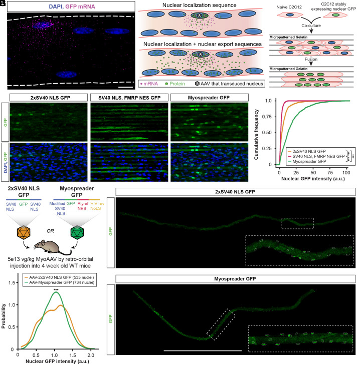
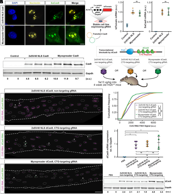
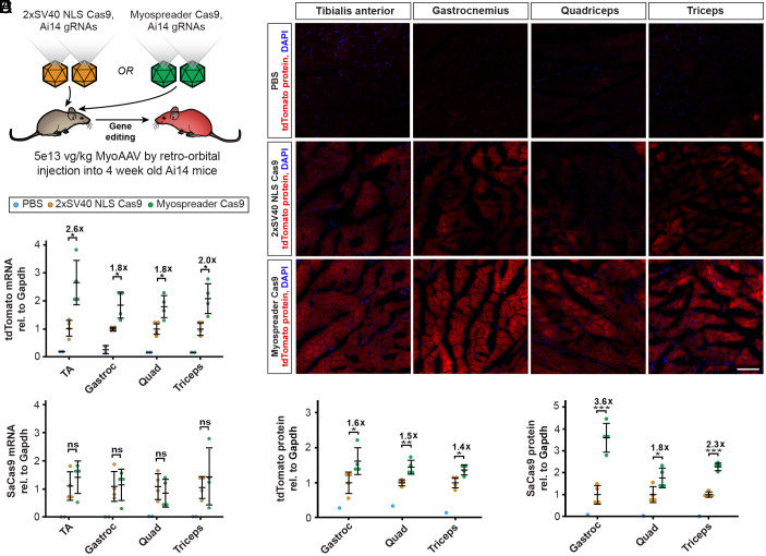
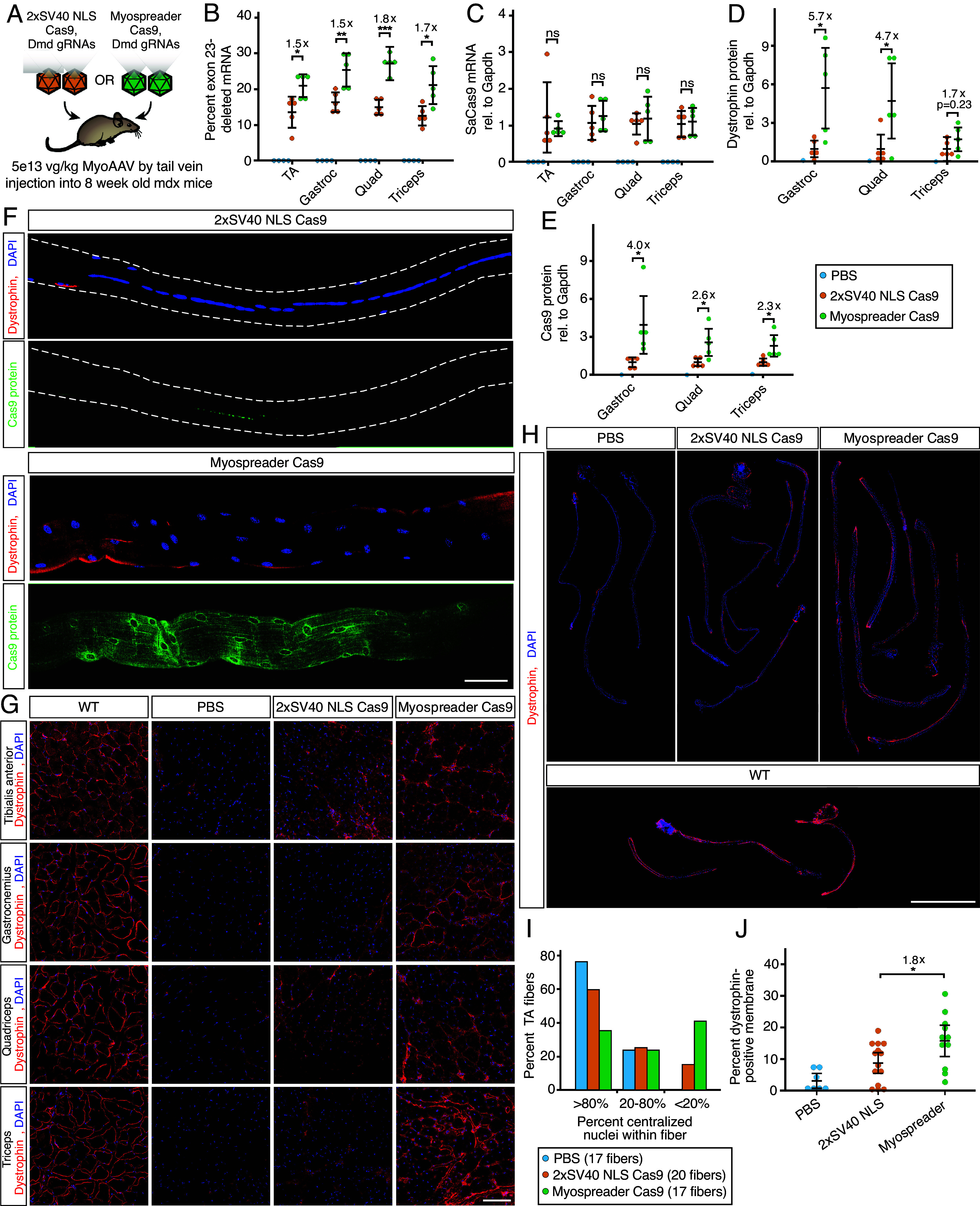
Successful CRISPR/Cas9-based gene editing in skeletal muscle is dependent on efficient propagation of Cas9 to all myonuclei in the myofiber. However, nuclear-targeted gene therapy cargos are strongly restricted to their myonuclear domain of origin. By screening nuclear localization signals and nuclear export signals, we identify "Myospreader," a combination of short peptide sequences that promotes myonuclear propagation. Appending Myospreader to Cas9 enhances protein stability and myonuclear propagation in myoblasts and myofibers. AAV-delivered Myospreader dCas9 better inhibits transcription of toxic RNA in a myotonic dystrophy mouse model. Furthermore, Myospreader Cas9 achieves higher rates of gene editing in CRISPR reporter and Duchenne muscular dystrophy mouse models. Myospreader reveals design principles relevant to all nuclear-targeted gene therapies and highlights the importance of the spatial dimension in therapeutic development.

Keywords: CRISPR; adeno-associated virus; gene therapy; myonuclear domains; spatial gene regulation.


Conflict of interest statement

Competing interests statement:E.T.W. is a co-founder and consultant to Kate Therapeutics. K.K.P. and E.T.W. are inventors on a patent related to this work.

Figures

Fig. 1.
Fig. 1.
Nuclear import and export sequence combinations enhance myonuclear GFP trafficking in myotubes and myofibers. (A) HCR-FISH against GFP mRNA (magenta) in a tibialis anterior (TA) myofiber of a mouse treated with an AAV encoding GFP. (Scale bar: 10 µm.) (B) Model for why protein cargoes with nuclear localization sequences accumulate in certain nuclei and not others. Peptide tags that facilitate both nuclear import and export may improve trafficking of protein cargoes to nontransduced nuclei. (C) Schematic of the C2C12 fusion experiment to identify NLS/NES combinations that improve propagation of GFP across multiple myonuclei. (D) Representative images of GFP fluorescence (green) in chimeric C2C12 myotubes expressing NLS/NES combinations. (Scale bar: 40 µm.) (E) Cumulative distribution functions of nuclear GFP signal for NLS/NES combinations in chimeric C2C12 myotubes. Significance by Kolmogorov–Smirnov test. (F) Schematic of the experiment to assess nuclear propagation of GFP in 4-wk-old WT mice treated systemically with 5E+13vg/kg 2xSV40 NLS GFP AAV or Myospreader GFP AAV. (G) GFP signal (green) in representative TA myofibers isolated from treated mice. Myonuclei borders are indicated with dashed lines. (Scale bar: 1 mm.) (H) Density plot of nuclear GFP signal in TA myofibers of treated mice. Significance by Kolmogorov–Smirnov test (ns = not significant; *P < 0.05; **P < 0.01; ***P < 0.001) (error bars = 95% CI).
Fig. 2.
Fig. 2.
Myospreader improves stability and localization of Cas9/dCas9 in myoblasts and myofibers. (A) Representative IF images of C2C12 myoblasts transfected with plasmids encoding 2xSV40 Cas9 or Myospreader Cas9. SaCas9 is shown in green, nucleolin in yellow, and DAPI in blue. (Scale bars: 10 µm.) (B) Schematic of the experiment to assess Myospreader Cas9 stability and editing efficiency in C2C12 myoblasts. (C) tdTomato mRNA expression as assessed by RT-qPCR in AI14 reporter C2C12s following transfection with plasmids encoding 2xSV40 Cas9 or Myospreader Cas9. Plotted relative to the mean of the 2xSV40 NLS Cas9 group. Significance by Student’s t test. (D) Cas9 mRNA expression as assessed by RT-qPCR in AI14 reporter C2C12s following transfection with plasmids encoding 2xSV40 Cas9 or Myospreader Cas9. Plotted relative to the mean of the 2xSV40 NLS Cas9 group. Significance by Student’s t test. (E) Western blot against Cas9 and Gapdh in Ai14 reporter C2C12s following transfection with plasmids encoding 2xSV40 Cas9 or Myospreader Cas9. Relative signal intensity determined by densitometry at the bottom. A.U.: arbitrary unit, normalized to Gapdh. (F) Schematic of dCas9 impeding transcription of toxic CUG RNA and experiment. Each treatment was packaged in a single AAV vector systemically delivered to 4-wk-old HSALR mice at a dose of 5E+13 vg/kg. (G) HCR-FISH to detect CUG RNA foci (magenta) and dCas9 mRNA (green) in myonuclei of representative TA myofibers isolated from treated HSALR mice. Myonuclei borders are indicated with dashed lines. Asterisks indicate dCas9-positive myonuclei. (Scale bar: 30 µm.) (H) Cumulative distribution functions of HCR-FISH CUG RNA signal in myonuclei proximal to dCas9-expressing myonuclei in myofibers isolated from treated HSALR mice. Significance by KolmogorovSmirnov test. (I) dCas9 mRNA expression as assessed by RT-qPCR in gastrocnemius muscle of treated HSALR mice. Plotted relative to the mean of the 2xSV40 NLS Cas9 nontargeting group. Significance by Tukey’s HSD. (J) Western blot detecting dCas9 and Gapdh in gastrocnemius muscle of treated mice. Relative signal intensity determined by densitometry at the bottom. A.U.: arbitrary unit, normalized to Gapdh (ns = not significant; *P < 0.05; **P < 0.01; ***P < 0.001) (error bars = 95% CI).
Fig. 3.
Fig. 3.
Myospreader improves Cas9 editing efficiency in vivo. (A) Schematic of the experiment to measure the efficacy of AAVs containing 2xSV40 NLS Cas9 or Myospreader Cas9, systemically delivered to 4-wk-old Ai14 mice at a dose of 5E+13 vg/kg. (B) tdTomato mRNA expression as assessed by RT-qPCR in muscles of treated Ai14 mice. Plotted relative to the mean of the 2xSV40 NLS Cas9 group. Significance by Student’s t test. (C) Cas9 mRNA expression as assessed by RT-qPCR in muscles of treated Ai14 mice. Plotted relative to the mean of the 2xSV40 NLS Cas9 group. Significance by Student’s t test. (D) Representative images of tdTomato fluorescence (red) and DAPI (blue) in muscles of treated Ai14 mice. (Scale bar: 100 µm.) (E) Quantification of Tdtomato protein as determined by western blot/densitometry, normalized to Gapdh. Plotted relative to the mean of the 2xSV40 NLS Cas9 group. Significance by Student’s t test. (F) Quantification of Cas9 protein as determined by western blot/densitometry, normalized to Gapdh. Plotted relative to the mean of the 2xSV40 NLS Cas9 group. Significance by Student’s t test (ns = not significant; *P < 0.05; **P < 0.01; ***P < 0.001) (error bars = 95% CI).
Fig. 4.
Fig. 4.
Myospreader improves Cas9 editing efficiency in a Mouse model of Duchenne muscular dystrophy. (A) Schematic of the experiment to measure the efficacy of AAVs containing 2xSV40 NLS Cas9 or Myospreader Cas9, systemically delivered to 4-wk-old mdx mice at a dose of 5E+13 vg/kg. (B) RT-qPCR quantification of exon-23 deleted Dmd mRNA in different muscles of treated mdx mice. Significance by Student’s t test. (C) RT-qPCR quantification of Cas9 mRNA in different muscles of treated mdx mice. Plotted relative to the mean of the 2xSV40 NLS Cas9 group. Significance by Student’s t test. (D) Quantification of dystrophin protein as determined by western blot/densitometry, normalized to Gapdh. Plotted relative to the mean of the 2xSV40 NLS Cas9 group. Significance by Student’s t test. (E) Quantification of Cas9 protein as determined by western blot/densitometry, normalized to Gapdh. Plotted relative to the mean of the 2xSV40 NLS Cas9 group. Significance by Student’s t test. (F) IF to detect dystrophin (red) and Cas9 (green) protein in representative TA myofibers isolated from treated mdx mice. (Scale bar: 50 µm.) (G) Representative images of Dystrophin protein (red) and DAPI (blue) in muscle cryosections of treated mdx mice by IF. (Scale bar: 100 µm.) (H) IF to detect dystrophin protein (red) and DAPI (blue) in whole TA myofibers isolated from treated mdx mice or untreated 8-wk-old WT mice. (Scale bar: 1 mm.) (I) Quantification of centralized nuclei in TA myofibers isolated from treated mdx mice. (J) Quantification of dystrophin protein at the periphery of TA myofibers of treated mdx mice or 8-wk-old WT mice. Significance determined Student’s t test (ns = not significant; *P < 0.05; **P < 0.01; ***P < 0.001) (error bars = 95% CI).

Update of

References

    1. Denes L. T., Kelley C. P., Wang E. T., Microtubule-based transport is essential to distribute RNA and nascent protein in skeletal muscle. Nat. Commun. 12, 6079 (2021). - PMC - PubMed
    1. Merlie J. P., Sanes J. R., Concentration of acetylcholine receptor mRNA in synaptic regions of adult muscle fibres. Nature 317, 66–68 (1985). - PubMed
    1. Fontaine B., Sassoon D., Buckingham M., Changeux J. P., Detection of the nicotinic acetylcholine receptor alpha-subunit mRNA by in situ hybridization at neuromuscular junctions of 15-day-old chick striated muscles. EMBO J. 7, 603–609 (1988). - PMC - PubMed
    1. Pavlath G. K., Rich K., Webster S. G., h. Blau M., Localization of muscle gene products in nuclear domains. Nature 337, 570–573 (1989). - PubMed
    1. Morin A., et al. , Dystrophin myonuclear domain restoration governs treatment efficacy in dystrophic muscle. Proc. Natl. Acad. Sci. U.S.A. 120, e2206324120 (2023). - PMC - PubMed

Publication types

Substances

LinkOut - more resources