

Box A of HMGB1 Maintains the DNA Gap and Prevents DDR-induced Kidney Injury in D-galactose Induction Rats
- PMID: 38688613
- PMCID: PMC11059889
- DOI: 10.21873/invivo.13552
Box A of HMGB1 Maintains the DNA Gap and Prevents DDR-induced Kidney Injury in D-galactose Induction Rats
Abstract
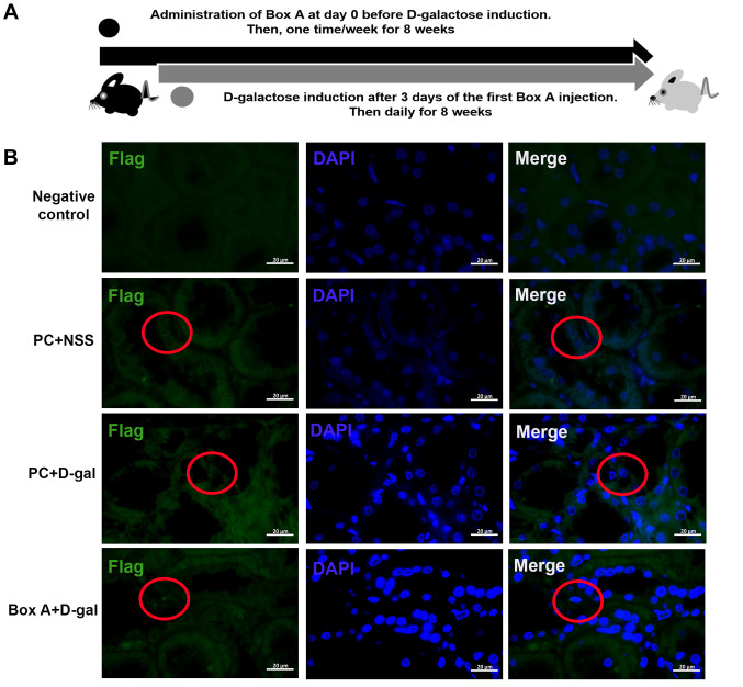
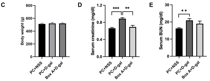
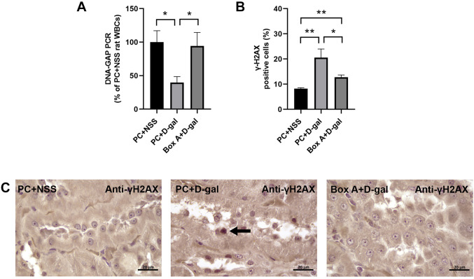
Background/aim: Disability and mortality rates for renal failure are still increasing. DNA damage and oxidative stress intoxication from body metabolism, high blood glucose, or the environment cause significant kidney damage. Recently, we reported that Box A of HMGB1 (Box A) acts as molecular scissors, producing DNA gaps that prevent DNA damage in kidney cell lines and ultimately reverse aging phenotypes in aging rat models. The present study aimed to demonstrate the potency of Box A in preventing D-galactose (D-gal)-induced kidney injury.
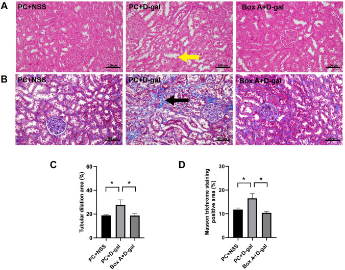
Materials and methods: A Box A expression plasmid was constructed and administered to a rat model. D-gal was injected subcutaneously for eight weeks. Serum was collected to study renal function, and white blood cells were collected for DNA gap measurement. Kidney tissue was also collected for γ-H2AX and NF-κB immunostaining; Senescence-associated (SA)-beta-gal staining; and analysis of the mRNA expression of p16INK4A, TNF-α, and IL-6. Moreover, histopathology analysis was performed using hematoxylin & eosin and Masson trichome staining.
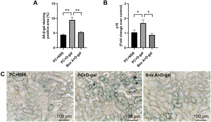
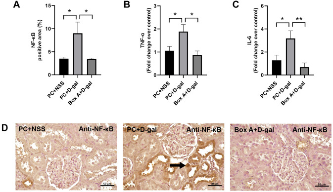
Results: Pretreatment with Box A administration prevented the reduction of DNA gaps and the consequences of the DNA damage response, which include elevated serum creatinine; high serum BUN; an increased positive SA-beta-gal staining area; overexpression of p16INK4A, NF-κB and senescence-associated secretory phenotype molecules, including IL-6, TNF-α; and histological alterations, including tubular dilation and collagen accumulation.
Conclusion: Box A effectively prevents DNA gap reduction and all D-gal-induced kidney pathological changes at the molecular, histological, and physiological levels. Therefore, Box A administration is a promising novel therapeutic strategy to prevent DNA-damaging agent-induced kidney failure.
Keywords: Box A of HMGB1; DNA damage; DNA durability; DNA protection; Kidney failure; youth-DNA gap.
Copyright © 2024, International Institute of Anticancer Research (Dr. George J. Delinasios), All rights reserved.
Conflict of interest statement
The Authors declare that there are no conflicts of interest in relation to this study.
Figures
Similar articles
-
Gensenoside Rg1 protects against lipopolysaccharide- and d-galactose-induced acute liver failure via suppressing HMGB1-mediated TLR4-NF-κB pathway.Mol Cell Probes. 2021 Apr;56:101706. doi: 10.1016/j.mcp.2021.101706. Epub 2021 Feb 20. Mol Cell Probes. 2021. PMID: 33617946
-
The neurovascular protective effects of huperzine A on D-galactose-induced inflammatory damage in the rat hippocampus.Gerontology. 2014;60(5):424-39. doi: 10.1159/000358235. Epub 2014 Jun 21. Gerontology. 2014. PMID: 24969491
-
Mechanism of ginsenoside Rg1 renal protection in a mouse model of d-galactose-induced subacute damage.Pharm Biol. 2016 Sep;54(9):1815-21. doi: 10.3109/13880209.2015.1129543. Epub 2016 Jan 5. Pharm Biol. 2016. PMID: 26730750
-
A high-fat diet increases oxidative renal injury and protein glycation in D-galactose-induced aging rats and its prevention by Korea red ginseng.J Nutr Sci Vitaminol (Tokyo). 2014;60(3):159-66. doi: 10.3177/jnsv.60.159. J Nutr Sci Vitaminol (Tokyo). 2014. PMID: 25078371
-
Emerging role of NF-κB signaling in the induction of senescence-associated secretory phenotype (SASP).Cell Signal. 2012 Apr;24(4):835-45. doi: 10.1016/j.cellsig.2011.12.006. Epub 2011 Dec 11. Cell Signal. 2012. PMID: 22182507 Review.
Cited by
-
Imipramine-induced Apoptosis and Metastasis Inhibition in Human Bladder Cancer T24 Cells Through EGFR/ERK/NF-κB Pathway Suppression.In Vivo. 2025 Mar-Apr;39(2):669-682. doi: 10.21873/invivo.13871. In Vivo. 2025. PMID: 40010952 Free PMC article.
-
The role of Box A of HMGB1 in producing γH2AX associated DNA breaks in lung cancer.Sci Rep. 2025 Jan 25;15(1):3215. doi: 10.1038/s41598-025-87773-3. Sci Rep. 2025. PMID: 39863746 Free PMC article.
-
HMGB1-BoxA gene therapy in reversing cisplatin resistance in non-small cell lung cancer.PLoS One. 2025 Jun 25;20(6):e0327144. doi: 10.1371/journal.pone.0327144. eCollection 2025. PLoS One. 2025. PMID: 40561066 Free PMC article.
-
Long-term Safety Pharmacology and Musculoskeletal Changes of HMGB1 Box A Gene Therapy in Middle-aged Monkeys.In Vivo. 2025 Jul-Aug;39(4):1965-1983. doi: 10.21873/invivo.13994. In Vivo. 2025. PMID: 40579035 Free PMC article.
References
MeSH terms
Substances
LinkOut - more resources
Full Text Sources
Molecular Biology Databases
Miscellaneous