Clinical Trial
. 2024 Jun;30(6):1602-1611.
doi: 10.1038/s41591-024-02965-0. Epub 2024 Apr 30.

Neoadjuvant nivolumab with or without relatlimab in resectable non-small-cell lung cancer: a randomized phase 2 trial

Affiliations
Clinical Trial

Neoadjuvant nivolumab with or without relatlimab in resectable non-small-cell lung cancer: a randomized phase 2 trial

Martin Schuler et al. Nat Med. 2024 Jun.

Abstract

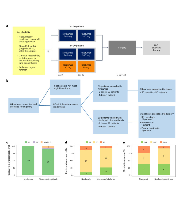
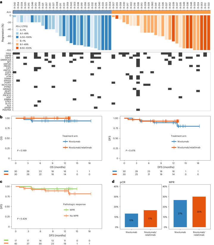
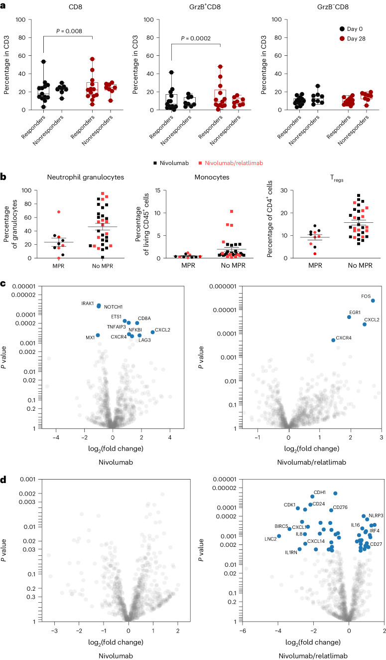
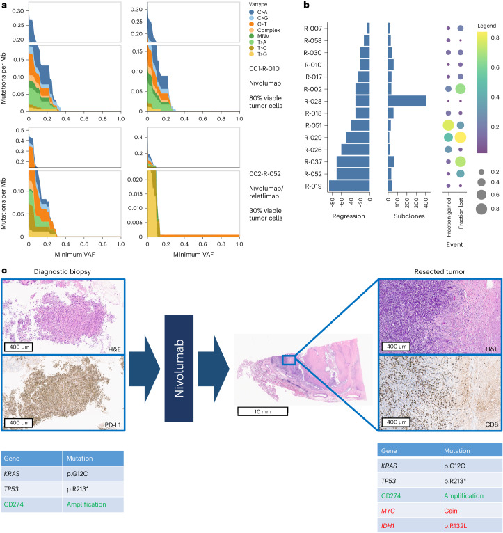
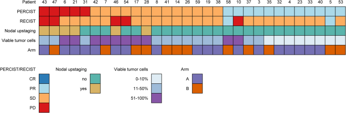
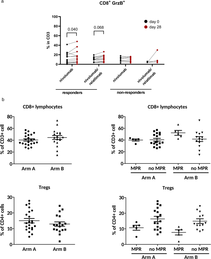
Antibodies targeting the immune checkpoint molecules PD-1, PD-L1 and CTLA-4, administered alone or in combination with chemotherapy, are the standard of care in most patients with metastatic non-small-cell lung cancers. When given before curative surgery, tumor responses and improved event-free survival are achieved. New antibody combinations may be more efficacious and tolerable. In an ongoing, open-label phase 2 study, 60 biomarker-unselected, treatment-naive patients with resectable non-small-cell lung cancer were randomized to receive two preoperative doses of nivolumab (anti-PD-1) with or without relatlimab (anti-LAG-3) antibody therapy. The primary study endpoint was the feasibility of surgery within 43 days, which was met by all patients. Curative resection was achieved in 95% of patients. Secondary endpoints included pathological and radiographic response rates, pathologically complete resection rates, disease-free and overall survival rates, and safety. Major pathological (≤10% viable tumor cells) and objective radiographic responses were achieved in 27% and 10% (nivolumab) and in 30% and 27% (nivolumab and relatlimab) of patients, respectively. In 100% (nivolumab) and 90% (nivolumab and relatlimab) of patients, tumors and lymph nodes were pathologically completely resected. With 12 months median duration of follow-up, disease-free survival and overall survival rates at 12 months were 89% and 93% (nivolumab), and 93% and 100% (nivolumab and relatlimab). Both treatments were safe with grade ≥3 treatment-emergent adverse events reported in 10% and 13% of patients per study arm. Exploratory analyses provided insights into biological processes triggered by preoperative immunotherapy. This study establishes the feasibility and safety of dual targeting of PD-1 and LAG-3 before lung cancer surgery.ClinicalTrials.gov Indentifier: NCT04205552 .


Conflict of interest statement

M.S., K.C., B.H., J.K., F.M., A.P., B.M., H.R., P.B., A.S. and C.A. received institutional funding, paid to the University Hospital Essen, from Bristol Myers Squibb to support the study NEOpredict-Lung. The following authors declare potential conflicts of interest: M.S. (Fees for consulting from Amgen, AstraZeneca, Blueprint Medicines, Boehringer Ingelheim, Bristol Myers Squibb, GSK, Janssen, MSD, Novartis, Roche, Sanofi and Tacalyx; honoraria for CME presentations from Amgen, Bristol Myers Squibb, GSK, Janssen, MSD, Roche and Sanofi; institutional research funding to University Hospital Essen from AstraZeneca, Bristol Myers Squibb and Janssen); K.C. (Fees for consulting from AstraZeneca/Medimmune, Bayer, Bristol Myers Squibb, MSD; institutional research funding from Bristol Myers Squibb; travel support from MSD; other relationship to Bristol Myers Squibb); M.W. (Fees for consulting from Daiichi Sankyo, Janssen, Novartis, Pfizer and Roche; honoraria for CME presentations from Amgen, AstraZeneca, GSK, Janssen, Novartis, Roche and Takeda; institutional research funding to University Hospital Essen from Bristol Myers Squibb and Takeda; travel support from Amgen and Bristol Myers Squibb), B.H. (institutional research funding to University Hospital Essen from Bristol Myers Squibb), K.D. (Fees for consulting from bess, Boehringer Ingelheim, Boston Scientific, Broncus Medical, FreeFlow, Fujifilm, Lys Medical, Medtronic, Morair Medtech, Olympus, Pulmonx and Storz; honoraria for CME presentations from AstraZeneca, Boehringer Ingelheim, Boston Scientific, Broncus Medical, Erbe Elektromedizin, Olympus and Storz; institutional research funding to University Medicine Essen – Ruhrlandklinik from Ambu, Broncus Medical, Epigenomics, Gala Therapeutics, Novartis, Nuvaira, PneumRx, Pulmonx and Roche); H.H. (Honoraria for CME presentations from Urenco; travel support from Pari; institutional research funding to University Hospital Essen from Pari), H.R. (Honoraria for CME presentations from AstraZeneca, Bristol Myers Squibb, Boehringer Ingelheim, Chop GmbH, Diaceutics, GSK, HUeG, Janssen-Cilag, MCI, Merck, Novartis, Roche Pharma, Sanofi and Wolfsburg Klinikum; institutional research funding from Bristol Myers Squibb), P.B. (Fees for consulting from AstraZeneca, Beigene, Bristol Myers Squibb, Merck Sharp & Dohme; honoraria from Bristol Myers Squibb, MSD; research funding to the Netherlands Cancer Institute from Bristol Myers Squibb; travel support from MSD; other relationship to Bristol Myers Squibb), A.S. (Institutional research funding to University Hospital Essen from Bristol Myers Squibb) and C.A. (Fees for consulting from Ewimed; honoraria for CME presentations from AstraZeneca, Bristol Myers Squibb, Roche; institutional research funding to University Hospital Essen Bristol Myers Squibb, and to University Medicine Essen – Ruhrlandklinik from PharmaCept). The other authors declare no competing interests.

Figures

Fig. 1
Fig. 1. Study design, patient deposition and secondary endpoints.
a, Graphical representation of clinical study design including key inclusion criteria. b, Patient deposition during the phases of the clinical study including screening, preoperative immunotherapy and curative resection. Reasons for screening failure and outcomes of surgery are summarized (*including one patient with single bone metastasis). c, Fraction of patients (n = 60) with microscopically complete (R0, green), microscopically incomplete (R1, purple) and macroscopically incomplete (pleural carcinosis, M1a (PLE), orange) resection of primary lung cancers and, if present, lymph node metastases per study arm. d, Fraction of patients (n = 60) with complete (none), partial response (PR, green), stable (SD, yellow) and progressive disease (PD, red) per RECIST evaluation of CT scans per study arm. e, Fraction of patients (n = 31) with complete (none), partial metabolic response (PMR, green), metabolically stable (SMD, yellow) and metabolically progressive disease (PMD, red) per PERCIST evaluation of positron emission tomography scans per study arm. SoC, standard of care.
Fig. 2
Fig. 2. Pathological responses, biomarkers and survival outcomes.
a, Waterfall plots of pathologic tumor regression (percentage reduction of viable tumor cells) in resected tumors and lymph nodes following neoadjuvant treatment with nivolumab (arm A, blue) or nivolumab and relatlimab (arm B, red). The color intensity encodes the category of PD-L1 expression by tumor cells (TPS <1% light color, TPS 1–49% medium dark color, TPS 50–100% dark color). The lower panel depicts the oncogram of each tumor using next-generation DNA sequencing of 500 cancer-related genes. Boxes represent pathogenic genomic aberrations in the respective gene. Genes with pathogenic aberrations in at least two study patients are listed. b, Kaplan–Meier plots for OS (left) and DFS (right) per study arm (arm A nivolumab, blue; arm B nivolumab and relatlimab, red). c, Kaplan–Meier plot for DFS in patients achieving a MPR (≤10% viable tumor cells (green)), and not achieving a MPR (>10% viable tumor cells (orange)). Statistical comparisons by log-rank test, vertical lines indicate censored patients. Two patients (both arm A) had died from noncancer causes. Six patients (four in arm A and two in arm B) have recurred or died. No patient with MPR has recurred, one patient with MPR had died from a noncancer cause. d, Fraction and number of patients with complete pathological response (pCR, upper) and MPR (lower) in study arms A (nivolumab (blue)) and B (nivolumab and relatlimab (red)).
Fig. 3
Fig. 3. Immune cell subsets and gene expression in peripheral blood and resected tumors.
a, Fraction of total CD8+ T cells (left), CD8+GrzB+ effector T cells (center) and CD8+GrzB T cells (right) in the peripheral blood of responding (≤50% viable tumor cells in resected tumors and lymph nodes) and nonresponding patients (>50% viable tumor cells). Each dot represents an individual patient: baseline values are in black and values at day 28 are in red. Whiskers and boxes represent the minimum, first, second and third quartiles and the maximum. Wilcoxon matched pairs signed-rank test was applied for statistical comparison. All P values are two-sided, no adjustments were made for multiple comparisons. b, Fraction of CD16+ neutrophil granulocytes (left), CD14+ monocytes (center) and CD4+CD25+ regulatory T cells (Treg, right) in single-cell suspensions from resected tumors. Each dot or box represents a single patient (black, nivolumab; red, nivolumab and relatlimab; MPR, ≤10% viable tumor cells in resected tumors and lymph nodes; no MPR, >10% viable tumor cells). Horizontal lines indicate the mean and s.e.m. c, Differential expression of immune-related and cancer pathway-related genes in response to treatment with nivolumab (left) and nivolumab and relatlimab (right) are presented as volcano plots. Significantly (FDR ≤ 0.05) upregulated (right of 0 line on x axes) and downregulated (left of 0 line on x axes) genes are depicted as blue closed circles. Selected significantly regulated genes are indicated. P values on the y axes were calculated using the two-sided quasi-likelihood F-test approach of EdgeR. d, Differential expression of immune-related genes and cancer pathway-related genes in resected tumors with MPR following treatment with nivolumab (left) and nivolumab and relatlimab (right) compared with resected tumors without MPR. Significantly (FDR ≤ 0.05) upregulated (right of 0 line on x axes) and downregulated (left of 0 line on x axes) genes in tumors with MPR are depicted as blue closed circles. Selected significantly regulated genes are indicated. P values on the y axes were calculated using the two-sided quasi-likelihood F-test approach of EdgeR. There was no significant interaction with MPR following nivolumab treatment.
Fig. 4
Fig. 4. Dynamic changes in the mutational spectra in response to immunotherapy.
a, Prevalence of mutations per megabase (Mb, y axes) of 500 cancer-related genes in pretherapeutic diagnostic biopsies (left) and resected tumors (right) of two exemplary patients without (001-R-010) and with response (002-R-052) to study therapy. The specific mutations (nucleotide exchanges from C to A (C>A), G (C>G) or T (C>T), from T to A (T>A), C (T>C) or G (T>G), complex nucleotide replacements (complex) or multiple nucleotide variants (MNV)) are color-coded from dark blue to yellow. The minimal VAFs are depicted on the x axes. b, Subclonal dynamics between pretherapeutic biopsies and resected tumors of 14 patients; each line depicts an individual patient. Left, pathological regression (percentage reduction of viable tumor cells) following immunotherapy. Center, estimated total number of subclones in the resected tumor. Right, fraction of subclones enriched (‘fraction gained’) and depleted (‘fraction lost’) in the resected tumors. Fractions are visualized by color (with yellow for high, purple for low), and bubble size (large for high, small for low, no bubble for zero). c, Selection of genomically encoded putative resistance mechanisms in one of 43 patients with pretreatment and posttreatment tumor specimens for genomic analyses. Left, representative microphotographs of the pretherapeutic diagnostic tumor biopsy stained with H&E and with an anti-PD-L1 primary antibody. DNA sequencing of the tumor biopsy revealed pathogenic mutations of KRAS and TP53 and amplification of the CD274 (PD-L1)-encoding gene. Center, low magnification image of a H&E-stained section of the resected tumor showing massive necrosis, but a residual region of vital tumor cells on the left-hand margin. Right, high magnification photomicrographs representing the transition zone from necrotic tumor to residual viable tumor cells stained with H&E and with an anti-CD8 primary antibody demonstrating tumor-infiltrating T lymphocytes. DNA sequencing of the resected tumor confirmed the presence of pathogenic mutations of KRAS and TP53 and amplification of the CD274 (PD-L1)-encoding gene. In addition, copy number gain of MYC and a pathogenic IDH1 mutation were newly detected. A complete list of patients with enrichment of genomically encoded putative resistance mechanisms in resected tumors is presented in Supplementary Fig. 3.
Extended Data Fig. 1
Extended Data Fig. 1. Response assessment per FDG-PET/CT and CT in relation to pathological response and nodal upstaging.
Metabolic responses (PERCIST), radiographic responses (RECIST), nodal upstaging (yes, no), pathological response category (Viable tumor cells), and treatment arm (A – nivolumab, B – nivolumab/relatlimab) of 30 patients from study site Essen, who underwent FDG-PET/CT scanning following preoperative immunotherapy. One patient was excluded because surgery was aborted due to pleural carcinosis.
Extended Data Fig. 2
Extended Data Fig. 2. Immune cell subsets in peripheral blood and resected tumors.
a, Induction of CD8+GrzB+ effector T cells in the peripheral blood in response to neoadjuvant nivolumab or nivolumab and relatlimab treatment. Each dot represents an individual patient, base line values in black, values at day 28 (after neoadjuvant immunotherapy) in red. Responders are defined by ≤ 50% viable tumor cells in resected tumors and lymph nodes, non-responders by >50% viable tumor cells. The Wilcoxon matched pairs signed-rank test was applied for statistical comparison. All p-values are two-sided, no adjustment was made for multiple comparisons. b, Characterization of infiltrating T lymphocytes in resected tumors in relation to study treatment (arm A: nivolumab, arm B: nivolumab and relatlimab) and achieving a major pathological response (MPR, ≤ 10% viable tumor cells in resected tumors and lymph nodes) or not achieving a MPR (no MPR). Each symbol represents an individual patient. Tregs – regulatory T lymphocytes. Horizontal lines indicate the mean value and standard error of the mean.

Comment in

References

    1. Sung H, et al. Global Cancer Statistics 2020: GLOBOCAN estimates of incidence and mortality worldwide for 36 cancers in 185 countries. CA Cancer J. Clin. 2021;71:209–249. - PubMed
    1. Reck M, Remon J, Hellmann MD. First-line immunotherapy for non-small-cell lung cancer. J. Clin. Oncol. 2022;40:586–597. - PubMed
    1. Tan AC, Tan DSW. Targeted therapies for lung cancer patients with oncogenic driver molecular alterations. J. Clin. Oncol. 2022;40:611–625. - PubMed
    1. Tsuboi M, et al. Overall survival with osimertinib in resected EGFR-mutated NSCLC. N. Engl. J. Med. 2023;389:137–147. - PubMed
    1. Solomon BJ, et al. LBA2 ALINA: efficacy and safety of adjuvant alectinib versus chemotherapy in patients with early-stage ALK+ non-small cell lung cancer (NSCLC) Ann. Oncol. 2023;34:S1295–S1296.

Publication types

MeSH terms

Associated data