. 2024 May;56(5):1137-1149.
doi: 10.1038/s12276-024-01221-2. Epub 2024 May 1.

Targeting pyruvate dehydrogenase kinase 1 overcomes EGFR C797S mutation-driven osimertinib resistance in non-small cell lung cancer

Affiliations

Targeting pyruvate dehydrogenase kinase 1 overcomes EGFR C797S mutation-driven osimertinib resistance in non-small cell lung cancer

Wonyoung Park et al. Exp Mol Med. 2024 May.

Abstract

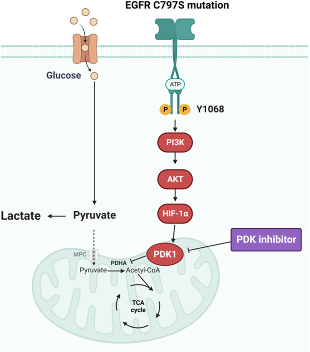
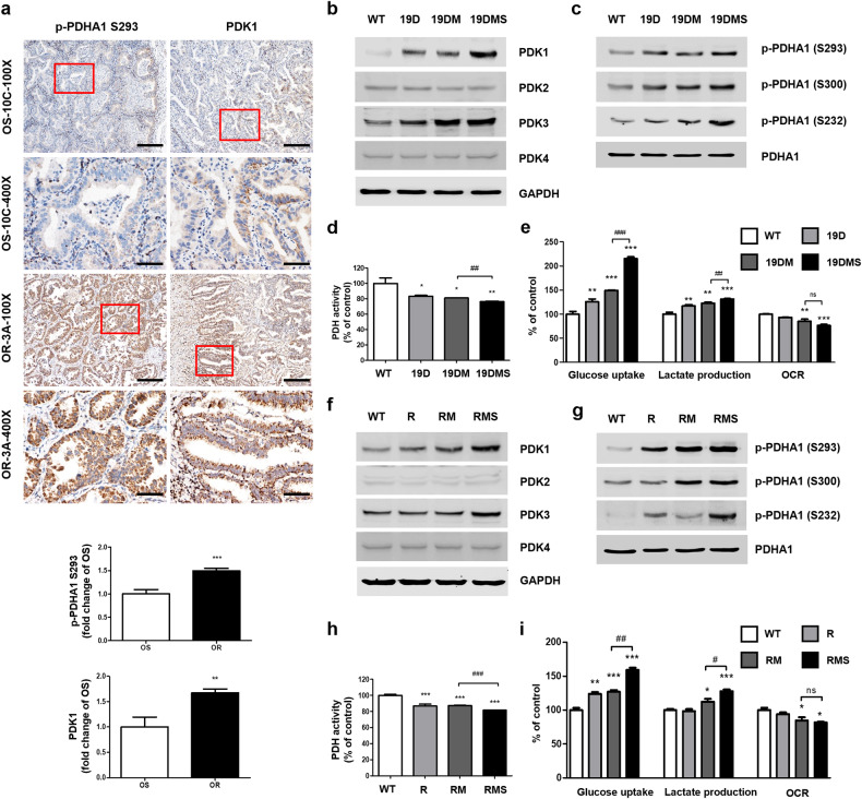
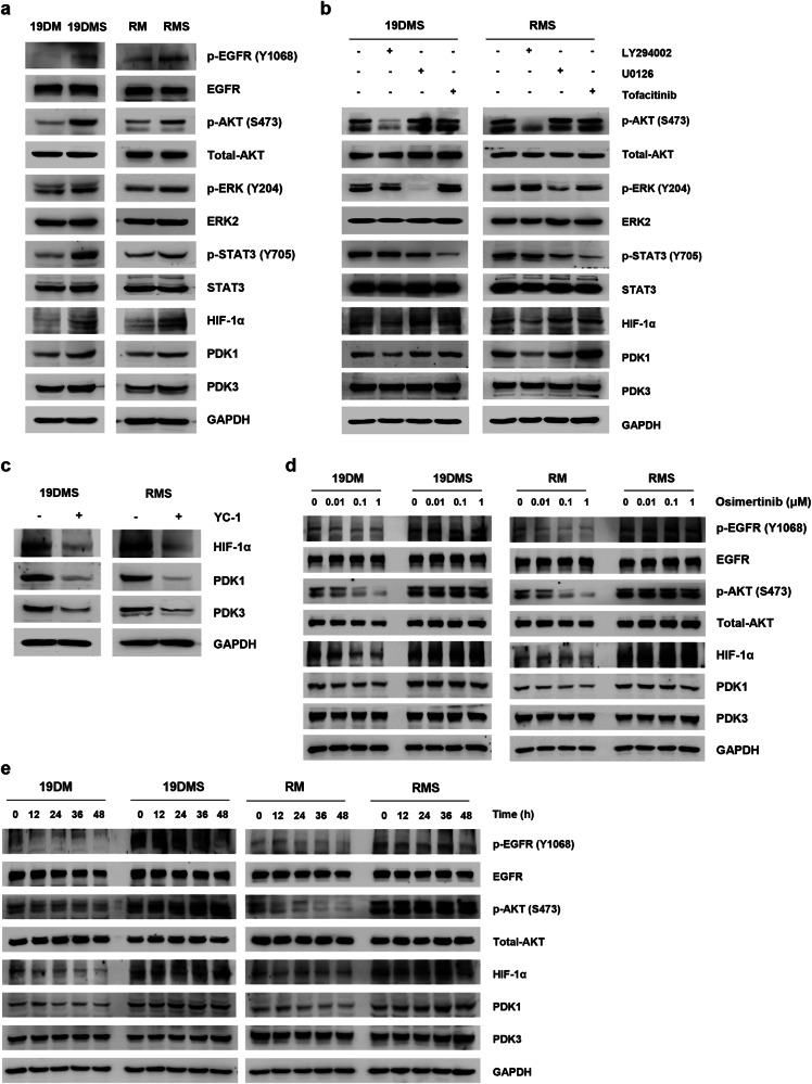
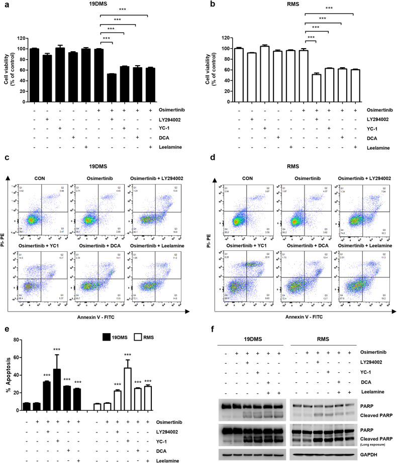
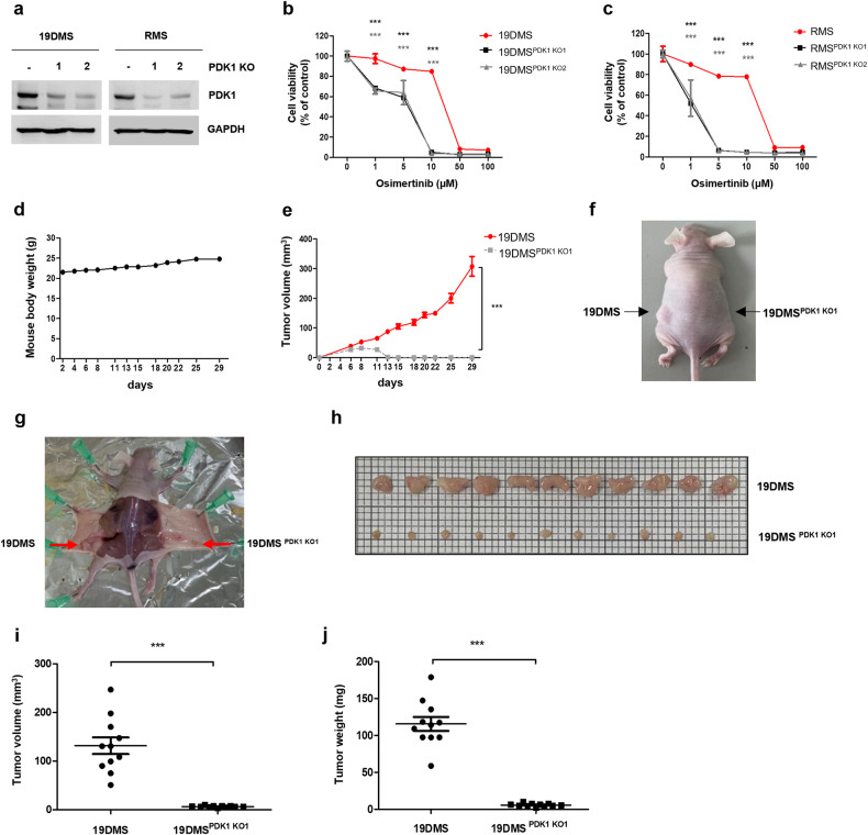
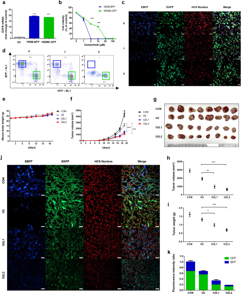
Osimertinib, a selective third-generation epidermal growth factor receptor (EGFR) tyrosine kinase inhibitor (TKI), effectively targets the EGFR T790M mutant in non-small cell lung cancer (NSCLC). However, the newly identified EGFR C797S mutation confers resistance to osimertinib. In this study, we explored the role of pyruvate dehydrogenase kinase 1 (PDK1) in osimertinib resistance. Patients exhibiting osimertinib resistance initially displayed elevated PDK1 expression. Osimertinib-resistant cell lines with the EGFR C797S mutation were established using A549, NCI-H292, PC-9, and NCI-H1975 NSCLC cells for both in vitro and in vivo investigations. These EGFR C797S mutant cells exhibited heightened phosphorylation of EGFR, leading to the activation of downstream oncogenic pathways. The EGFR C797S mutation appeared to increase PDK1-driven glycolysis through the EGFR/AKT/HIF-1α axis. Combining osimertinib with the PDK1 inhibitor leelamine helped successfully overcome osimertinib resistance in allograft models. CRISPR-mediated PDK1 knockout effectively inhibited tumor formation in xenograft models. Our study established a clear link between the EGFR C797S mutation and elevated PDK1 expression, opening new avenues for the discovery of targeted therapies and improving our understanding of the roles of EGFR mutations in cancer progression.


Conflict of interest statement

The authors declare no competing interests.

Figures

Fig. 1
Fig. 1. Schematic diagram showing the mechanism of osimertinib resistance driven by the EGFR C797S mutation.
The schematic illustrates the mechanism of epidermal growth factor receptor (EGFR) C797S mutation-driven osimertinib resistance due to the increase in pyruvate dehydrogenase kinase (PDK)1 expression through the EGFR/Serine/Threonine Kinase 1 (AKT)/hypoxia-inducible factor (HIF)-1α axis; the combination of osimertinib with a PDK inhibitor can overcome this resistance.
Fig. 2
Fig. 2. Osimertinib-resistant non-small cell lung cancers (NSCLCs) exhibit elevated PDK1 expression, followed by enhanced glycolysis.
a Representative immunohistochemical images showing phosphorylated pyruvate dehydrogenase E1 subunit alpha 1 (p-PDHA1) (S293) and pyruvate dehydrogenase kinase 1 (PDK1) in lung cancer tissues of who are osimertinib-sensitive (OS) and osimertinib-resistant (OR) patients. The area in the red box represents a view at 400× magnification. The scale bar represents 200 μm at 100× magnification and 50 μm at 400× magnification. b, f The expression levels of PDK1, PDK2, PDK3, PDK4, and glyceraldehyde 3-phosphate dehydrogenase (GAPDH; loading control) in A549 epidermal growth factor receptor (EGFR) wild-type and mutant cells were measured via western blotting. c, g p-PDHA1 (S293), p-PDHA1 (S300), p-PDHA1 (S232), and total PDHA1 levels in A549 EGFR wild-type and mutant cells were measured via western blotting. d, h PDH activity was measured in A549 EGFR wild-type and mutant cells. e, i Glucose uptake, lactate production, and oxygen consumption rate (OCR) were measured in A549 EGFR wild-type and mutant cells. Data information: The data in a, d, e, h, and i are presented as the mean ± standard error of the mean (SEM) values. Statistical analysis (a, d, e, h and i) was conducted using Student’s t-test for comparisons among the OS, EGFR19del747_750+T790M (19DM), and EGFRL858R+T790M (RM) groups. Statistical analyses in d, e, h, and i were performed using one-way analysis of variance with Dunnett’s post hoc test, with comparison to the wild-type (WT) group. *p < 0.05, **p < 0.01, ***p < 0.001, #p < 0.05, ##p < 0.01, and ###p < 0.001 indicate statistically significant differences. “ns” indicates nonsignificance.
Fig. 3
Fig. 3. The epidermal growth factor receptor (EGFR) C797S mutation results in PDK1 upregulation via the EGFR/AKT/HIF-1α axis in A549 cells.
a Immunoblot analysis results showing the levels of p-EGFR (Y1068), p-AKT Serine/Threonine Kinase 1 (AKT) (S473), p-extracellular signal-regulated kinase (ERK) (Y204), and p-signal transducer and activator of transcription 3 (STAT3) (Y705), as well as the levels of EGFR, total AKT, ERK2, STAT3, hypoxia-inducible factor (HIF)-1α, pyruvate dehydrogenase kinase (PDK)1, PDK3, and glyceraldehyde 3-phosphate dehydrogenase (GAPDH; loading control) in EGFR-mutant A549 cells. b Immunoblot analysis of A549 EGFR19del747_750+T790M+C797S (19DMS)/EGFRL858R+T790M+C797S (RMS) cells treated with LY294002 (50 μM), U0126 (10 μM), or tofacitinib (50 μM) for 24 h. The levels of p-AKT (S473), p-ERK (Y204), and p-STAT3 (Y705), as well as the levels of total AKT, ERK2, STAT3, HIF-1α, PDK1, PDK3, and GAPDH (loading control), were measured. c Immunoblot analysis of A549 19DMS/RMS cells treated with YC-1 (50 μM) for 24 h. The expression of HIF-1α, PDK1, PDK3, and GAPDH (loading control) was measured. d Immunoblot analysis of A549 19DMS/RMS cells treated with the indicated concentrations of osimertinib for 24 h. The levels of p-EGFR (Y1068) and p-AKT (S473), as well as the levels of EGFR, total AKT, HIF-1α, PDK1, PDK3, and GAPDH (loading control), were measured. e Immunoblot analysis of A549 19DMS/RMS cells treated with osimertinib (0.1 μM) at the indicated time points. The levels of p-EGFR (Y1068) and p-AKT (S473), as well as the levels of EGFR, total AKT, HIF-1α, PDK1, PDK3, and GAPDH (loading control), were measured.
Fig. 4
Fig. 4. The combination of pyruvate dehydrogenase kinase (PDK) inhibitors and osimertinib amplifies apoptosis in A549 epidermal growth factor receptor (EGFR) C797S mutant cells.
A549 EGFR19del747_750+T790M+C797S (19DMS)/EGFRL858R+T790M+C797S (RMS) cells were treated with osimertinib (1 μM) alone or in combination with LY294002 (50 μM), YC-1 (50 μM), DCA (15 mM), or leelamine (3 μM) for 24 h. a, b Cell viability was assessed using the 3-(4,5-dimethylthiazol-2-yl)-2,5-diphenyl-2H-tetrazolium bromide (MTT) assay. c-e Apoptotic cells were detected using Annexin V/PI staining and flow cytometry; flow cytometry-based dot plots of the Annexin V/PI assay are shown. f Western blotting was performed to evaluate the levels of poly (ADP-ribose) polymerase (PARP), cleaved PARP, and glyceraldehyde 3-phosphate dehydrogenase (GAPDH; loading control). Data information: The data in a, b, and e are presented as the mean ± standard error of the mean (SEM) values. Statistical analyses in a and b were conducted using a one-way analysis of variance with Dunnett’s post hoc test, with comparison to the osimertinib treatment group (sixth lane). Statistical analysis in e was performed using one-way analysis of variance with Dunnett’s post hoc test, with comparison to the control group (the first lane for 19DMS or RMS cells). ***p < 0.001.
Fig. 5
Fig. 5. Combined targeting of PDK1 inhibits xenograft tumor formation and increases osimertinib sensitivity in A549 EGFR C797S mutant cells.
a Pyruvate dehydrogenase kinase (PDK)1 sgRNA was transfected into A549 EGFR19del747_750+T790M+C797S (19DMS)/EGFRL858R+T790M+C797S (RMS) cells, and PDK1 and glyceraldehyde 3-phosphate dehydrogenase (GAPDH; loading control) expression was measured by immunoblotting. b Treatment of A549 EGFR19del747_750+T790M+C797S (19DMS), 19DMSPDK1 KO1, and 19DMSPDK1 KO2 cells with osimertinib for 72 h, followed by measurement of cell viability using the 3-(4,5-dimethylthiazol-2-yl)-2,5-diphenyl-2H-tetrazolium bromide (MTT) assay. c Treatment of A549 EGFRL858R+T790M+C797S (RMS), RMSPDK1 KO1, and RMSPDK1 KO2 cells with osimertinib for 72 h, followed by the measurement of cell viability using the MTT assay. dj A549 19DMS (left) or A549 19DMSPDK1 KO1 (right) cells (1 × 107) suspended in 100 μL of sterile phosphate-buffered saline were injected subcutaneously into BALB/c nude mice. Mouse body weight (d) and tumor volume (e) were monitored. Representative images of mice (f, g) and tumors (h). Tumor volume (i) and tumor weight (j). Data information: The data in b, c, e, i, and j are presented as the mean ± standard error of the mean (SEM) values. Statistical analyses in b, c were conducted using Student’s t-test, with comparison to the control group (b, 19DMS; c, RMS). Statistical analyses in e, i, and j were performed using Student’s t-test, with comparison to the 19DMS group. ***p < 0.001.
Fig. 6
Fig. 6. The combination of osimertinib and leelamine effectively inhibits EGFR C797S mutant tumor growth in vivo.
a Confirmation of epidermal growth factor receptor (EGFR) mutant (EGFR19del747_750+T790M (19DM)-blue fluorescent protein (BFP)) or EGFR19del747_750+T790M+C797S (19DMS)-green fluorescent protein (GFP) overexpression in LLC cells using qPCR analysis of EGFR mRNA expression. b Assessment of the cytotoxic effects of osimertinib on EGFR-mutant LLC cells (19DM-BFP or 19DMS-GFP) using the 3-(4,5-dimethylthiazol-2-yl)-2,5-diphenyl-2H-tetrazolium bromide (MTT) assay after treatment with the indicated concentrations of osimertinib for 72 h. c Fluorescence microscopy images showing a 1:1 mixture of EGFR-mutant 19DM-BFP and 19DMS-GFP cells treated with osimertinib (0, 1, 5 μM) for 72 h. The scale bar represents 50 μm. d Flow cytometry data confirming the ratio of BFP to GFP in LLC 19DM-BFP and LLC 19DMS-GFP cells mixed at a 1:1 ratio and treated with osimertinib (0, 1, 5 μM) for 72 h. ek Allograft models were established by injecting a 1:1 mixture of 19DM-BFP and LLC 19DMS-GFP LLC cells into C57BL/6 mice. After one week, the mice were treated with corn oil (CON), 2 mg/kg osimertinib (O2), 2 mg/kg osimertinib + 1 mg/kg leelamine (O2L1), or 2 mg/kg osimertinib + 2 mg/kg leelamine (O2L2) for two weeks. Body weight (e) and tumor volume (f) were measured during the treatment period. The mice were euthanized, tumor samples were harvested, and the tumors were imaged (g). Tumor volume (h) and weight (i) were also analyzed. Imaging of the tumors was performed using a confocal microscope (j). The scale bar represents 20 μm. The fluorescence intensity ratio of 19DM-BFP and 19DMS-GFP LLC cells was quantified using ImageJ software, and the bar length indicates the relative tumor volume across different treatment groups (k). Data information: The data in b, f, h, and i are presented as the mean ± standard error of the mean (SEM) values. Statistical analysis in b was conducted using Student’s t-test, with comparison to the 19DM group. Statistical analyses in f, h, and i were performed using a one-way analysis of variance with Dunnett’s post hoc test, with comparison to the O2 group. *p < 0.05, **p < 0.01, and ***p < 0.001.

References

    1. Herbst RS. Review of epidermal growth factor receptor biology. Int. J. Radiat. Oncol. Biol. Phys. 2004;59:21–26. doi: 10.1016/j.ijrobp.2003.11.041. - DOI - PubMed
    1. Ciardiello F, Tortora G. EGFR antagonists in cancer treatment. N. Engl. J. Med. 2008;358:1160–1174. doi: 10.1056/NEJMra0707704. - DOI - PubMed
    1. Zhang YL, et al. The prevalence of EGFR mutation in patients with non-small cell lung cancer: a systematic review and meta-analysis. Oncotarget. 2016;7:78985–78993. doi: 10.18632/oncotarget.12587. - DOI - PMC - PubMed
    1. Sharma SV, Bell DW, Settleman J, Haber DA. Epidermal growth factor receptor mutations in lung cancer. Nat. Rev. Cancer. 2007;7:169–181. doi: 10.1038/nrc2088. - DOI - PubMed
    1. Morgillo F, Della Corte CM, Fasano M, Ciardiello F. Mechanisms of resistance to EGFR-targeted drugs: lung cancer. ESMO Open. 2016;1:e000060. doi: 10.1136/esmoopen-2016-000060. - DOI - PMC - PubMed

MeSH terms