Case Reports
. 2024 Jun;11(6):1579-1589.
doi: 10.1002/acn3.52075. Epub 2024 Apr 30.

Clinical heterogeneity within the ALS-FTD spectrum in a family with a homozygous optineurin mutation

Affiliations
Case Reports

Clinical heterogeneity within the ALS-FTD spectrum in a family with a homozygous optineurin mutation

Tandis Parvizi et al. Ann Clin Transl Neurol. 2024 Jun.

Abstract

Objective: Mutations in the gene encoding for optineurin (OPTN) have been reported in the context of different neurodegenerative diseases including the amyotrophic lateral sclerosis (ALS) and frontotemporal dementia (FTD) spectrum. Based on single case reports, neuropathological data in OPTN mutation carriers have revealed transactive response DNA-binding protein 43 kDa (TDP-43) pathology, in addition to accumulations of tau and alpha-synuclein. Herein, we present two siblings from a consanguineous family with a homozygous frameshift mutation in the OPTN gene and different clinical presentations.

Methods: Both affected siblings underwent (i) clinical, (ii) neurophysiological, (iii) neuropsychological, (iv) radiological, and (v) laboratory examinations, and (vi) whole-exome sequencing (WES). Postmortem histopathological examination was conducted in the index patient, who deceased at the age of 41.

Results: The index patient developed rapidly progressing clinical features of upper and lower motor neuron dysfunction as well as apathy and cognitive deterioration at the age of 41. Autopsy revealed an ALS-FTLD pattern associated with prominent neuronal and oligodendroglial TDP-43 pathology, and an atypical limbic 4-repeat tau pathology reminiscent of argyrophilic grain disease. The brother of the index patient exhibited behavioral changes and mnestic deficits at the age of 38 and was diagnosed with behavioral FTD 5 years later, without any evidence of motor neuron dysfunction. WES revealed a homozygous frameshift mutation in the OPTN gene in both siblings (NM_001008212.2: c.1078_1079del; p.Lys360ValfsTer18).

Interpretation: OPTN mutations can be associated with extensive TDP-43 pathology and limbic-predominant tauopathy and present with a heterogeneous clinical phenotype within the ALS-FTD spectrum within the same family.


Conflict of interest statement

The authors declare that the research was conducted in the absence of any commercial or financial relationships that could be construed as a potential conflict of interest.

Figures

Figure 1
Figure 1
Cranial MRI revealing increased symmetrical T2 signal intensities along the pyramidal tracts (A) and motor band sign in SWI (B) in the index patient. The brother of the index patient showed a global gray matter volume loss (C) with no signs of paramagnetic susceptibility artifacts in the precentral gyrus (D).
Figure 2
Figure 2
Family pedigree depicting the clinical status. Genetic analysis was performed on the index patient (marked with an arrow) and the brother of the index patient. The pedigree included three cousins on the father's side diagnosed with slow‐progressing ALS. Cousin I was diagnosed at the age of 45 and died at the age of 55, cousin II was diagnosed at the age of 40 and died at the age of 54, and cousin III was diagnosed at the age of 43 and was admitted to a care home at the age of 48. An uncle on the mother's side developed dementia in his eighties and died at the age of 83, and another uncle on the father's side was also affected by dementia.
Figure 3
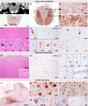
Figure 3
Neuropathological findings in the index patient. (A) MR image of the medulla oblongata confronted with a histological cross section showing both the prominent alteration of the pyramids with bright hyperintense signal in MR (upper panel) and prominent microglial / macrophage activation on immunohistochemistry (brown signal; lower panel); here, also microglial activation at the level of the XIIth cranial nerve and formatio reticularis is observed (immunohistochemistry for HLA‐DR). (B) Cross section through the thoracic spinal cord also reveals prominent microglial/macrophage activation along the lateral and anterior corticospinal tract and anterior horns of the spinal cord (immunohistochemistry for HLA‐DR). (C) Higher magnification comparing posterior (upper panel) and lateral columns (lower panel). (D) Neuronal loss at the anterior horn of the spinal cord (upper panel) and morphological changes of residual neurons with nearly normal appearance (left), shrunken perikaryon with condensed Nissl substance (second picture, arrow), condensation of cytoskeletal structures (third picture, arrowhead), and development of axonal swellings (right, empty arrowhead) and neuronophagia with macrophage clusters (right, arrow) (hematoxylin–eosin staining). (E) Immunohistochemistry for phospho‐TDP‐43 reveals abundant cytoplasmic aggregates in motor neurons ranging from a diffuse‐granular cytoplasmic staining to more skein‐like or fibrillar and more compact appearing inclusions (immunohistochemistry for phospho‐TDP‐43). (F) Oligodendroglial coiled body‐like inclusions are frequently observed in cortical (upper panel) and subcortical (left)/brainstem (center)/spinal cord (right) regions (immunohistochemistry for phospho‐TDP‐43). (G) Frontotemporal lobar degeneration pattern was also observed with superficial spongiosis in frontal and temporal cortices with neuronal loss and gliosis (hematoxylin–eosin staining). (H and I) In these areas, abundant neuronal and oligodendroglial pTDP‐43 inclusions are detected in all cortical layers (H) in form of a mainly diffuse‐granular or “dash‐like”‐pattern (I, arrows) with some coarser aggregates (I, arrowheads) and frequent oligodendroglial inclusions (I, empty arrowheads). In the hippocampus there is also moderate involvement of granule cells of the dentate gyrus (I, lower right). No obvious intranuclear inclusions are detected (immunohistochemistry for phospho‐TDP‐43). (J) In addition, there is prominent tau pathology limited to temporo‐medial regions (Immunohistochemistry for tau AT8). (K) There are frequent pretangles in all hippocampal sectors but particularly in CA2 (upper panel; immunohistochemistry for 4R‐tau), some ballooned neurons (lower left panel; immunohistochemistry for 4R‐tau), some grain‐like structures in the neuropil (lower right panel; immunohistochemistry for 4R‐tau) and (L) very abundant oligodendroglial inclusions in the white matter (upper panel; immunohistochemistry for 4R‐tau), and some granular fuzzy astrocytes in the gray matter (lower left panel, immunohistochemistry for 4R‐tau) all immunoreactive for 4‐repeat and negative for 3‐repeat tau isoforms (lower right panel; immunohistochemistry for 3R‐tau). Scale bars: 2 mm: A and J; 1 mm: B; 300 μm: G; 100 μm: D (upper panel) E (upper right), H; 50 μm: C (upper and lower panel), I (lower left & right panel); D; 20 μm: D (lower row), E (lower row & upper left), F and I (upper row & lower row center), K and L.
Figure 4
Figure 4
(A–C): Rt‐PCR revealed OPTN‐mRNA expression in PBMCs from the brother of the index patient (listed as Pat2, A). On a protein level, optineurin could not be detected in PMBC lysates (B) or lysates from postmortem frontal cortex of the index patient (listed as Pat1) in western blot analysis (C). (D–I) Anti‐phospho‐TDP‐43 immunohistochemistry on spinal cord sections of a sporadic case of ALS (D–F) and of the index patient ALS and the optineurin mutation (G–I). Phospho‐TDP43‐positive neuronal skein‐like and compact cytoplasmic inclusions in motor neurons in the control case (D) are also immunopositive (arrows) for two different anti‐optineurin antibodies (E: non‐C‐terminal; F: C‐terminal). In contrast, in the patient with the optineurin mutation, the inclusions (G) do not immunoreact with any of the two anti‐optineurin antibodies (H and I). Scale bars: 50 μm: D–F; 100 μm: G–I;

References

    1. Maruyama H, Morino H, Ito H, et al. Mutations of optineurin in amyotrophic lateral sclerosis. Nature. 2010;465(7295):223‐226. - PubMed
    1. Pottier C, Bieniek KF, Finch N, et al. Whole‐genome sequencing reveals important role for TBK1 and OPTN mutations in frontotemporal lobar degeneration without motor neuron disease. Acta Neuropathol. 2015;130(1):77‐92. - PMC - PubMed
    1. Dominguez J, Yu JT, Tan YJ, et al. Novel Optineurin frameshift insertion in a family with frontotemporal dementia and parkinsonism without amyotrophic lateral sclerosis. Front Neurol. 2021;12:645913. - PMC - PubMed
    1. Shen WC, Li HY, Chen GC, Chern Y, Tu PH. Mutations in the ubiquitin‐binding domain of OPTN/optineurin interfere with autophagy‐mediated degradation of misfolded proteins by a dominant‐negative mechanism. Autophagy. 2015;11(4):685‐700. - PMC - PubMed
    1. Nakazawa S, Oikawa D, Ishii R, et al. Linear ubiquitination is involved in the pathogenesis of optineurin‐associated amyotrophic lateral sclerosis. Nat Commun. 2016;7(1):12547. - PMC - PubMed

Publication types