Hemophilic pseudotumor of the knee joint: Emphasizing prevention and early diagnosis in a rare disease
- PMID: 38689692
- PMCID: PMC11059959
- DOI: 10.1002/ccr3.8822
Hemophilic pseudotumor of the knee joint: Emphasizing prevention and early diagnosis in a rare disease
Abstract
Key clinical message: Hemophilic pseudotumors are rare complications occurring in individuals with severe hemophilia, characterized by progressive cystic swellings in muscles and/or bones due to recurrent bleeding. Timely initiation of factor VIII replacement is crucial.
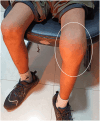
Abstract: Hemophilic pseudotumors are rare complications occurring in individuals with severe hemophilia, characterized by progressive cystic swellings in muscles and/or bones due to recurrent bleeding. Although their incidence has decreased with the advent of factor VIII replacement therapy, they still create challenges, particularly in regions with limited access to medical care. Here, we present a case report of a hemophilic pseudotumor of the knee joint in a 15-year-old male with hemophilia A. The patient presented with severe left knee pain, swelling, and restricted range of motion, prompting further investigation. Imaging studies revealed lytic lesions, and MRI bone signal changes consistent with hemophilic pseudotumors. Prompt initiation of factor VIII replacement therapy and supportive management led to a significant improvement in symptoms and joint functionality. Follow-up after 2 months showed that the swelling had significantly reduced in size, with marked improvement in the functionality of the knee joint. This case confirms what is already known in the hemophilia literature: how important it is to prevent, diagnose, and treat pseudotumors early in hemophilia. However, longer clinical and imaging follow-up of this case is necessary to determine whether the complaints associated with pseudotumors resolve with hematologic treatment or will require surgical treatment.
Keywords: hemophilia; hemophilic pseudotumor; knee joint; lytic bone lesion; multimodality imaging.
© 2024 The Authors. Clinical Case Reports published by John Wiley & Sons Ltd.
Conflict of interest statement
The authors have declared that no competing interests exist.
Figures


References
-
- Pakala A, Thomas J, Comp P. Hemophilic Pseudotumor: a case report and review of literature. Int J Clin Med. 2012;03(3):229‐233. doi: 10.4236/IJCM.2012.33046 - DOI
-
- Manco‐Johnson MJ, Soucie JM, Gill JC. Joint outcomes Committee of the Universal Data Collection, US hemophilia treatment center network. Prophylaxis usage, bleeding rates, and joint outcomes of hemophilia, 1999 to 2010: a surveillance project. Blood. 2017;129(17):2368‐2374. doi: 10.1182/blood-2016-02-683169 - DOI - PMC - PubMed
Publication types
LinkOut - more resources
Full Text Sources

