. 2024 Apr 16:15:1379376.
doi: 10.3389/fimmu.2024.1379376. eCollection 2024.

Tissue adaptation of CD4 T lymphocytes in homeostasis and cancer

Affiliations

Tissue adaptation of CD4 T lymphocytes in homeostasis and cancer

Marina V A Pereira et al. Front Immunol. .

Abstract

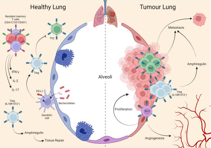
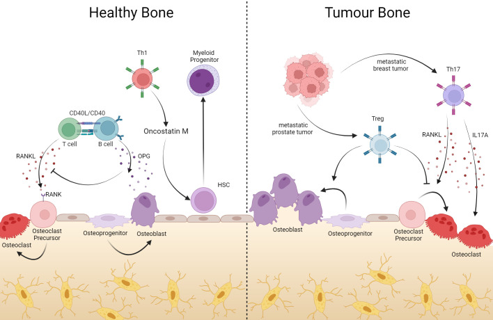
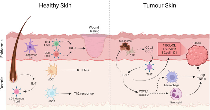
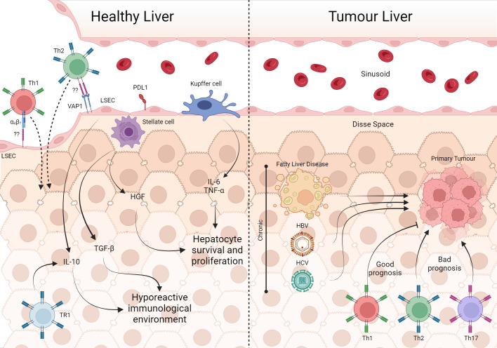
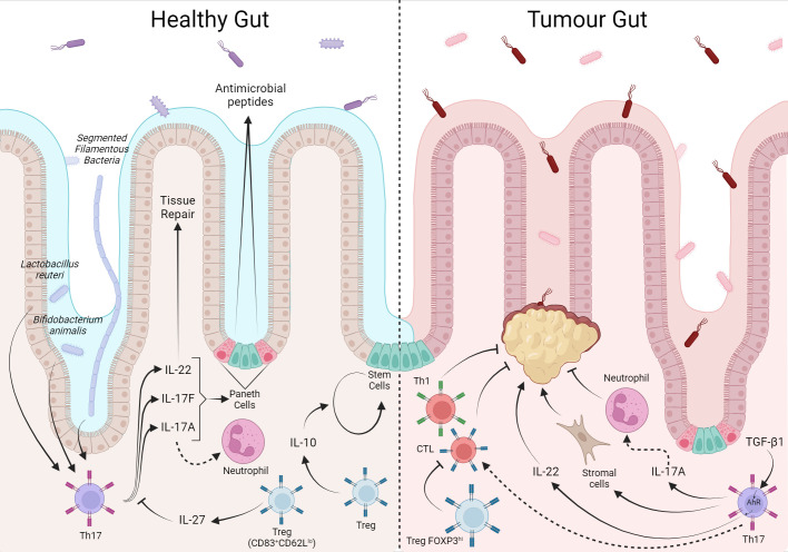
The immune system is traditionally classified as a defense system that can discriminate between self and non-self or dangerous and non-dangerous situations, unleashing a tolerogenic reaction or immune response. These activities are mainly coordinated by the interaction between innate and adaptive cells that act together to eliminate harmful stimuli and keep tissue healthy. However, healthy tissue is not always the end point of an immune response. Much evidence has been accumulated over the years, showing that the immune system has complex, diversified, and integrated functions that converge to maintaining tissue homeostasis, even in the absence of aggression, interacting with the tissue cells and allowing the functional maintenance of that tissue. One of the main cells known for their function in helping the immune response through the production of cytokines is CD4+ T lymphocytes. The cytokines produced by the different subtypes act not only on immune cells but also on tissue cells. Considering that tissues have specific mediators in their architecture, it is plausible that the presence and frequency of CD4+ T lymphocytes of specific subtypes (Th1, Th2, Th17, and others) maintain tissue homeostasis. In situations where homeostasis is disrupted, such as infections, allergies, inflammatory processes, and cancer, local CD4+ T lymphocytes respond to this disruption and, as in the healthy tissue, towards the equilibrium of tissue dynamics. CD4+ T lymphocytes can be manipulated by tumor cells to promote tumor development and metastasis, making them a prognostic factor in various types of cancer. Therefore, understanding the function of tissue-specific CD4+ T lymphocytes is essential in developing new strategies for treating tissue-specific diseases, as occurs in cancer. In this context, this article reviews the evidence for this hypothesis regarding the phenotypes and functions of CD4+ T lymphocytes and compares their contribution to maintaining tissue homeostasis in different organs in a steady state and during tumor progression.

Keywords: CD4 T lymphocytes; T CD4 tumor response; cancer; tissue-specific immune response; tumor immunology.


Conflict of interest statement

The authors declare that the research was conducted in the absence of any commercial or financial relationships that could be construed as a potential conflict of interest.

Figures

Figure 1
Figure 1
Interaction of T cells in lung homeostasis and cancer. During lung homeostasis in mice, the local microbiota induces PD-L1 in dendritic cells which support the generation of local Treg cells, important for suppressing the Th2 phenotype that is exacerbated in the tissue in the postnatal period. Another population of Treg (IL18R+/ST2+) assists tissue repair through the production of amphiregulin. In adulthood, mature human lung tissue is composed of memory T cells (CD4+ CD103+ CD69+) expressing IFN-ɣ, IL-2, and IL-17. These same phenotypes remain during the establishment of the tumor in human lung tissue, with emphasis on Th17 supporting tumor cell proliferation and angiogenesis. Mouse metastasis data demonstrate that Th2 and Treg (IL18R+/ST2+) are important for lung-specific metastasis in breast tumors. Created with BioRender.com.
Figure 2
Figure 2
In bone, T cells help with bone remodeling, but they can also support tumor processes. Bone resident T cells produce RANKL which, once bound to its RANK receptor on the surface of osteoclate precursors, induces the maturation of these cells, contributing to the composition of the bone matrix. Via CD40L-CD40 interaction, B cells produce OPG, inhibiting RANK-RANKL binding with consequent blockage of osteoclastogenesis. In addition to bone remodeling, CD4+ T lymphocytes act in hematopoiesis, where Oncotain M-producing Th1 cells induce HSC differentiation into myeloid progenitor. However, in the event of tumors or bone metastases, Th17 cells producing RANKL induce osteoclastogenesis, generating bone consumption in favor of the tumor. On the other hand, Treg cells can suppress differentiation into osteoclasts and assist in the differentiation of osteoblasts. Created with BioRender.com.
Figure 3
Figure 3
T cells in the skin play a role in wound healing and tumor pathogenesis. In healthy skin, Langerhan cells are the most abundant APC, besides that CD4+ and CD8+ T lymphocytes cells have an important role in wound healing producing IGF-1. On the other hand, memory T cells are in the dermis close to the follicle, an important producer of IL-7 that helps maintain the memory profile. Furthermore, dDC1 producing IFN-λ capable of cross-presentation and dDC2 important for Th2 generation. On the other hand, in tumor-affected skin Th17 cells expressing CXCL1 and CXCL2 contribute to the recruitment of macrophages and neutrophils producing IL-1β and TNF-α. Th17 directly through IL17 production induce expression of anti-apoptotic proteins and carcinogenic promoters such as Bcl-XL, Survivin, and Cyclin D1. Furthermore, cancer associated fibroblast cells produce CCL2 and CCL5, which are important for the recruitment of Th17 cells, contributing to the increase in this cell in the infiltrate. Created with BioRender.com.
Figure 4
Figure 4
The involvement of T cells in healthy liver and tumor processes. In the unique architecture of the liver, Kupffer cells, through IL-6 e TNF-α, are involved in the survival and proliferation of hepatocytes, as well as HGF from stellate cells. Furthermore, the production of IL-10 by TR1 and LSEC in addition to TGF-β from stellate cells maintains the hepatic microenvironment hyporesponsive. However, LSEC can recruit Th2 and Th1 via VAP-1 and α4 integrin, respectively which are important in infection contexts. Fatty liver disease, HBV and HCV are some of the risk factors for the development of liver tumors. With tumor establishment, cells from the Th1, Th2 and Th17 profiles are present in the tissue, with Th1 being associated with a good prognosis of the disease. Created with BioRender.com.
Figure 5
Figure 5
The T cell response in the intestine is balanced between tolerance to local microbiota and food antigens, but has a prognostic factor when in response to colorectal cancer. The local microbiota modulates the T cell phenotype, Lactobacillus reuteri and Bifidobacterium animalis induce the Th17 phenotype whose production of IL-22, 1L-17A/F, act in tissue repair, and induction of antimicrobial peptides production. IL-10 and IL-27 from Treg assist in tissue self-renewal and control of the local T response, respectively. In CRC, Th17 cells through IL17-A recruit antimoral neutrophils while they can also stimulate stromal cells in favor of tumor. Furthermore, under TGF-β1 stimulation from the tumor environment, IL-22 favors tumor growth. In contrast, local Treg inhibit tumor-inhibiting CD8+ and Th1.Created with BioRender.com.
Figure 6
Figure 6
Organ-specific signature based on the cytokine profile of CD4+ T cells. BALB/c female mice receive 5x10⁴ 4T1 cells inoculated subcutaneously in the fourth right mammary pad. On the days shown, animals were injected IV with anti-Thy1.2 mAb and three minutes later were euthanized. Cell suspension was obtained from each organ and stained for flow cytometry for CD4, TNF-α, IFN-y, IL-13, IL-17A, IL17-F and GM-CSF. Thy1.2+ cells were excluded from the analysis and the results shown were from gated CD4+ T lymphocytes. The mean fluorescence intensity of cytokines produced by CD4+ T lymphocytes from each organ was evaluated at two different moments of tumor progression; Day 17: tumor colonization in the breast draining lymph nodes, spleen, and lungs; Day 23: metastasis in all organs evaluated. (A) Principal component analysis (PCA) during tumor progression. Left panel: naïve (tumor free) PCA; Middle panel: Day 17 PCA; Right panel: Day 23 PCA. (B) PCA of each organ during tumor progression. Created with BioRender.com.

Similar articles

Cited by

References

    1. Eubank TD, Galloway M, Montague CM, Waldman WJ, Marsh CB. M-CSF induces vascular endothelial growth factor production and angiogenic activity from human monocytes. J Immunol. (2003) 171:2637–43. doi: 10.4049/jimmunol.171.5.2637 - DOI - PMC - PubMed
    1. Raggatt LJ, Wullschleger ME, Alexander KA, Wu ACK, Millard SM, Kaur S, et al. . Fracture healing via periosteal callus formation requires macrophages for both initiation and progression of early endochondral ossification. Am J Pathology. (2014) 184:3192–204. doi: 10.1016/j.ajpath.2014.08.017 - DOI - PubMed
    1. Shouval DS, Biswas A, Goettel JA, McCann K, Conaway E, Redhu NS, et al. . Interleukin-10 receptor signaling in innate immune cells regulates mucosal immune tolerance and anti-inflammatory macrophage function. Immunity. (2014) 40:706–19. doi: 10.1016/j.immuni.2014.03.011 - DOI - PMC - PubMed
    1. Ganusov VV, De Boer RJ. Do most lymphocytes in humans really reside in the gut? Trends Immunol. (2007) 28:514–8. doi: 10.1016/j.it.2007.08.009 - DOI - PubMed
    1. Smolders J, Remmerswaal EBM, Schuurman KG, Melief J, Van Eden CG, Van Lier RAW, et al. . Characteristics of differentiated CD8+ and CD4+ T cells present in the human brain. Acta Neuropathol. (2013) 126:525–35. doi: 10.1007/s00401-013-1155-0 - DOI - PubMed

Publication types