

Fracture Strength of Tooth Roots Following Canal Preparation by Three Rotary File Systems: An In Vitro Study
- PMID: 38690448
- PMCID: PMC11059113
- DOI: 10.7759/cureus.57302
Fracture Strength of Tooth Roots Following Canal Preparation by Three Rotary File Systems: An In Vitro Study
Abstract
Background: Since the beginning of modern endodontics, there have been many concepts, strategies, and techniques for root canal preparation. A mind-boggling variety of files have developed for negotiating and shaping them throughout the years. Today's most secure, most effective, and simplest file system combines the most reliable design elements of the past with the latest technological advances to create the most effective file system. So, the need for the study is to evaluate the fracture strength of tooth roots following canal preparation by three rotary file systems: ProTaper Universal file system (Dentsply, USA), ProTaper Next file system (Dentsply Sirona USA), and Neolix A1 nickel-titanium (NiTi) file system (Orikam Healthcare India Pvt Ltd., New Delhi, India).
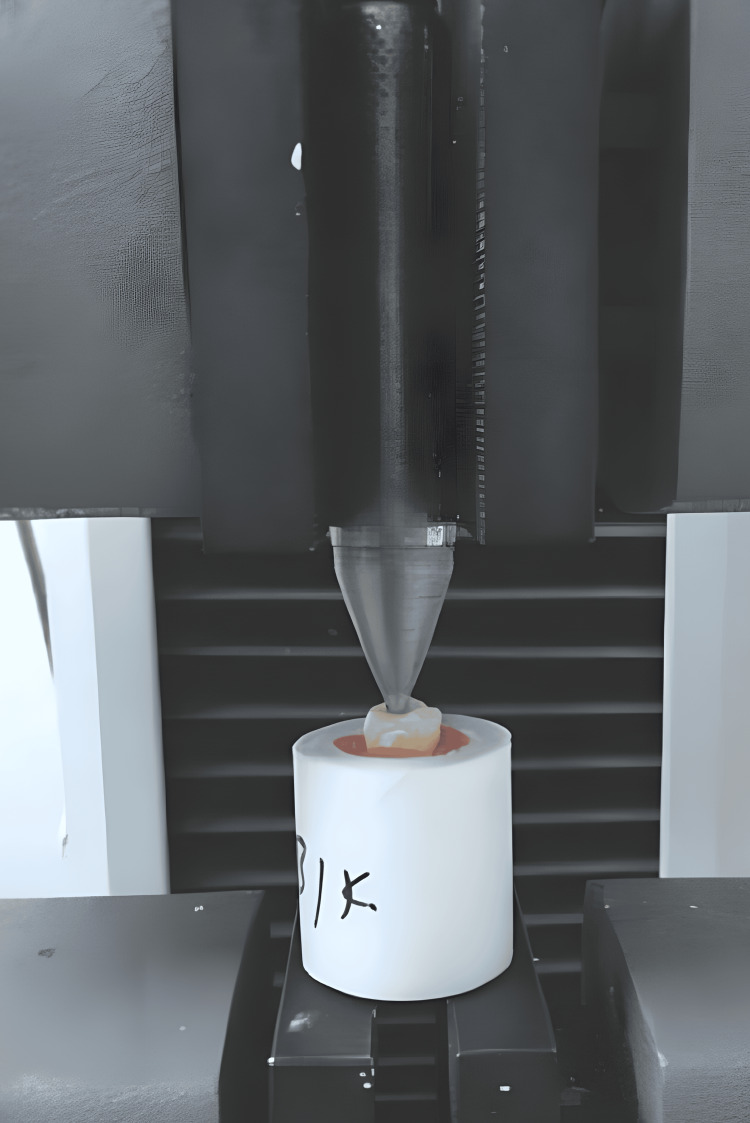
Method: Ninety human mandibular molars were selected for the study. Inclusion criteria include human mandibular first and second molars and teeth removed for routine clinical reasons, and intact apices were selected, excluding cases with root surface caries, root surface fissures, teeth with immature root apex, mesial canal fusion, extremely short roots, thin roots, or curved roots. All teeth were preserved in a solution of 10% neutral buffered formalin for two weeks and then transferred to distilled water for examination. The teeth were randomly divided into three groups. Access cavities were created, and working lengths were determined. Groups 1, 2, and 3 underwent shaping using ProTaper Universal, ProTaper Next, and Neolix A1 (NiTi) file systems, respectively, following guidelines. Canals were irrigated with sodium hypochlorite and ethylenediaminetetraacetic acid (EDTA) and were obturated up to the mid-root region with AH Plus sealer. To facilitate fracture testing, obturation was performed to distribute the load from the spreader to the canal wall. The EndoSequence and Quick-Fill obturation system were utilized to fill the apical half of the canal with gutta-percha material. After obturation, the distal root of each tooth was cut, while the mesial root was securely positioned in a putty material. A universal testing machine was employed for the fracture tests, operating at a cross-head speed of 1 mm/min. The machine was equipped with a D11 hand spreader tip, which was inserted into the root canal to make contact with the gutta-percha. Gradual force was applied to the root canal until a fracture occurred, at which point the force application was stopped. The amount of force required to cause the fracture was measured in newtons. Data were collected and recorded using IBM SPSS Statistics for Windows, Version 17.0 (Released 2008; IBM Corp., Armonk, New York, United States) and then transferred to Microsoft Excel for analysis. Descriptive statistics, mean, and standard deviation were used for continuous data. The fracture resistance of dental roots treated with three types of files was compared using a one-way ANOVA. Graphs were generated using Excel and Word. A significance level of p<0.01 was chosen.
Result: ANOVA indicated significant differences in mean fracture resistance: Neolix A1 (NiTi) (95.3 N) > NEXT (91.0 N) > universal (86.6 N), with a p-value of 0.004 (<0.001), confirming statistical significance.
Conclusion: The study concludes that the canal instrumented with Neolix A1 (NiTi) exhibits higher fracture resistance after canal instrumentation compared to ProTaper Next and ProTaper Universal.
Keywords: microcrack formation; neolix a1 file; protaper next; protaper universal; tooth fracture.
Copyright © 2024, Chiramel David et al.
Conflict of interest statement
The authors have declared that no competing interests exist.
Figures
References
-
- Endodontic complications of root canal therapy performed by dental students with stainless-steel K-files and nickel-titanium hand files. Pettiette MT, Metzger Z, Phillips C, Trope M. J Endod. 1999;25:230–234. - PubMed
-
- An in vitro comparison of incomplete root fractures associated with three obturation techniques. Onnink PA, Davis RD, Wayman BE. J Endod. 1994;20:32–37. - PubMed
-
- Effect of ethylenediaminetetraacetic acid on root fracture with respect to concentration at different time exposures. Uzunoglu E, Aktemur S, Uyanik MO, Durmaz V, Nagas E. J Endod. 2012;38:1110–1113. - PubMed
LinkOut - more resources
Full Text Sources
Miscellaneous