

Assessing the Accuracy of Lateral Cephalogram in Quantifying Three-Dimensional Pharyngeal Airway Morphology Compared to Cone-Beam Computed Tomography
- PMID: 38690459
- PMCID: PMC11059114
- DOI: 10.7759/cureus.57301
Assessing the Accuracy of Lateral Cephalogram in Quantifying Three-Dimensional Pharyngeal Airway Morphology Compared to Cone-Beam Computed Tomography
Abstract
Background: When it comes to orthodontic diagnosis and treatment planning, the structures of the upper and lower airway space are crucial because of the role they play in craniofacial development.
Aim: The major objective of this study was to evaluate the accuracy of lateral cephalogram in the evaluation of upper and lower pharyngeal space by comparing it to clinical usage of cone-beam computed tomography (CBCT) in quantifying the 3D morphology of the pharyngeal airway.
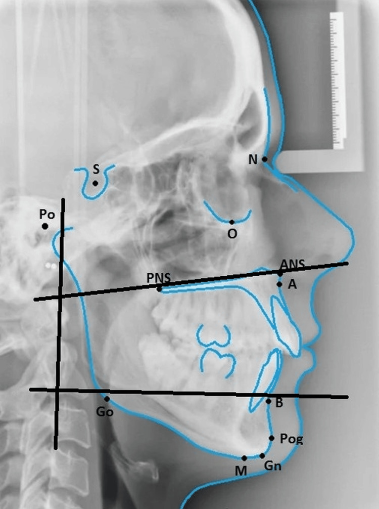
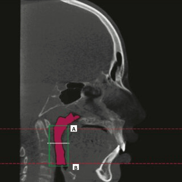
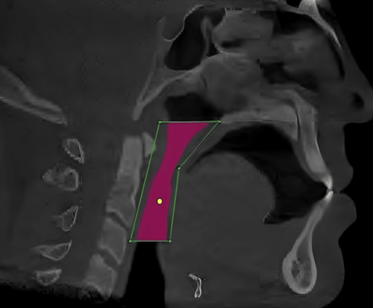
Methods and materials: In total, 70 patients were included in the study. They had both a CBCT scan and a lateral cephalogram performed within a week of each other. Different cephalometric landmarks have been utilized to estimate linear and area dimensions for use in lateral cephalogram airway investigations. By superimposing the lateral cephalogram measurement of the vertical height of the pharyngeal airway over axial CBCT slices of 0.8 to 1 mm in thickness, airway volumes were calculated. For this study, we measured the pharyngeal airway space in each patient in two dimensions (2D) using the airway area from the lateral cephalogram and in three dimensions (3D) using the airway volume from the CBCT scan over the same region of interest, using a uniform scale and magnification throughout all split 3D volumes.
Results: The mean value of the area of pharyngeal space calculated by lateral cephalograph analysis (LCA) was 336.35 ± 86.49 mm2. The maximum value was 551.234 mm2. The minimum value was 206.32 mm2. The mean value of the volume of the same area calculated using CBCT was 3409.11 ± 1237.96 mm3. The maximum value was 5887.23 mm3. When the area calculated using LCA was compared with the volume calculated using CBCT, the correlation between them was significant statistically (r=0.831, p-value =0.000). The mean values of volume evaluated in 3D CBCT in males were 4198±1008 mm3 while for females it was 2980±1134.5 mm3. During the statistical analysis, these observations were found to have a positive correlation with increased volume of pharyngeal space in males as compared to that of females (p=0.006). The values of the area of pharyngeal space calculated using LCA in males was 370.1±60.9 mm2. while it was 301.9±88 mm2 in females.
Conclusion: The area estimated for the pharyngeal airway on LCA correlates strongly with the volume determined by a CBCT scan. Since we have considered pharyngeal space analysis using CBCT to be a reliable and standard methodology, therefore a positive correlation of area calculated using LCA with volume calculated using CBCT shows that the analysis made by LCA can be reliable.
Keywords: cbct; lateral cephalogram; malocclusion; orthodontics; upper and lower pharyngeal space.
Copyright © 2024, Ashique Abdulhameed et al.
Conflict of interest statement
The authors have declared that no competing interests exist.
Figures
References
-
- Cone beam computed tomography: an innovative tool for airway assessment. Osorio F, Perilla M, Doyle DJ, Palomo JM. Anesth Analg. 2008;106:1803–1807. - PubMed
-
- Effects of mandibular advancement device (MAD) on airway dimensions assessed with cone-beam computed tomography. Haskell JA, McCrillis J, Haskell BS, Scheetz JP, Scarfe WC, Farman AG. Semin Orthod. 2009;15:132–158.
-
- Evaluation of pharyngeal airway space amongst different skeletal patterns. Alves M Jr, Franzotti ES, Baratieri C, Nunes LK, Nojima LI, Ruellas AC. Int J Oral Maxillofac Surg. 2012;41:814–819. - PubMed
-
- Evaluation of upper airway obstruction in Class II children with fluid-mechanical simulation. Iwasaki T, Saitoh I, Takemoto Y, Inada E, Kanomi R, Hayasaki H, Yamasaki Y. Am J Orthod Dentofacial Orthop. 2011;139:0–45. - PubMed
LinkOut - more resources
Full Text Sources
Research Materials